Stressor treatment networks
PDF
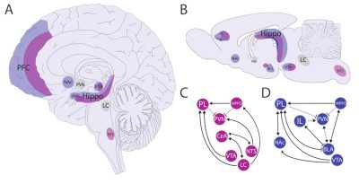
The detection and evaluation of different types of stressors (physical, in purple, and psychological, in blue) involve several structures, both in the human brain (A) and in that of rodents (B). The lower panels show that the processing of physical (C) and psychological (D) stressors requires the engagement of different networks (Figure 1).
Physical stressors primarily activate structures related to the control of vital functions located in the brainstem (in particular the nucleus of the tractus solitarius, NTS, and the locus coelucarius, LC) as well as the hypothalamus (in particular the paraventricular nucleus of the hypothalamus, PVN). However, prosencephalic regions are also involved in processing physical stressors, such as the prelimbic area (PL) of the prefrontal cortex (PFC). The central nucleus of the amygdala (CeA) is also involved in the integration of responses to physical stressors.
The prefrontal cortex is essential for developing appropriate responses to stressors, whether physical or psychological. It is strongly innervated by dopaminergic projections from the ventral tegmental area (VTA) and nucleus accumbens (NAc). Disruption of the prefrontal cortex is associated with anhedonia and aberrant reward-seeking behavior.
The integration of psychological stressors involves, in addition to the prelimbic area, another area of the prefrontal cortex, the infralimbic area (IL), as well as the basolateral nucleus of the amygdala (BLA).
The hippocampus (Hippo) is another brain structure activated in response to physical and psychological stressors. The CA1 region of the hippocampus has numerous connections with the limbic structures mentioned above, and the hippocampus is an important structure in the negative feedback of the corticotropic axis.
The paraventricular nucleus of the hypothalamus and the loecus cœruleus (PVN and LC, shown in grey) represent the main relays to the rest of the organism of the stress response, triggering the corticotropic axis and the autonomic nervous system respectively.


