| Focus 1/3 | Lichens, surprising pioneering organisms

Lichens: Hybrid organisms

PDF

Lichens are considered to be “cousins” of fungi because, traditionally, they are made up of a combination of a fungus and either a green algae or a cyanobacterium or both. This association is called symbiosis, the fungus is a mycosymbiote, and the algae a photosymbiote.

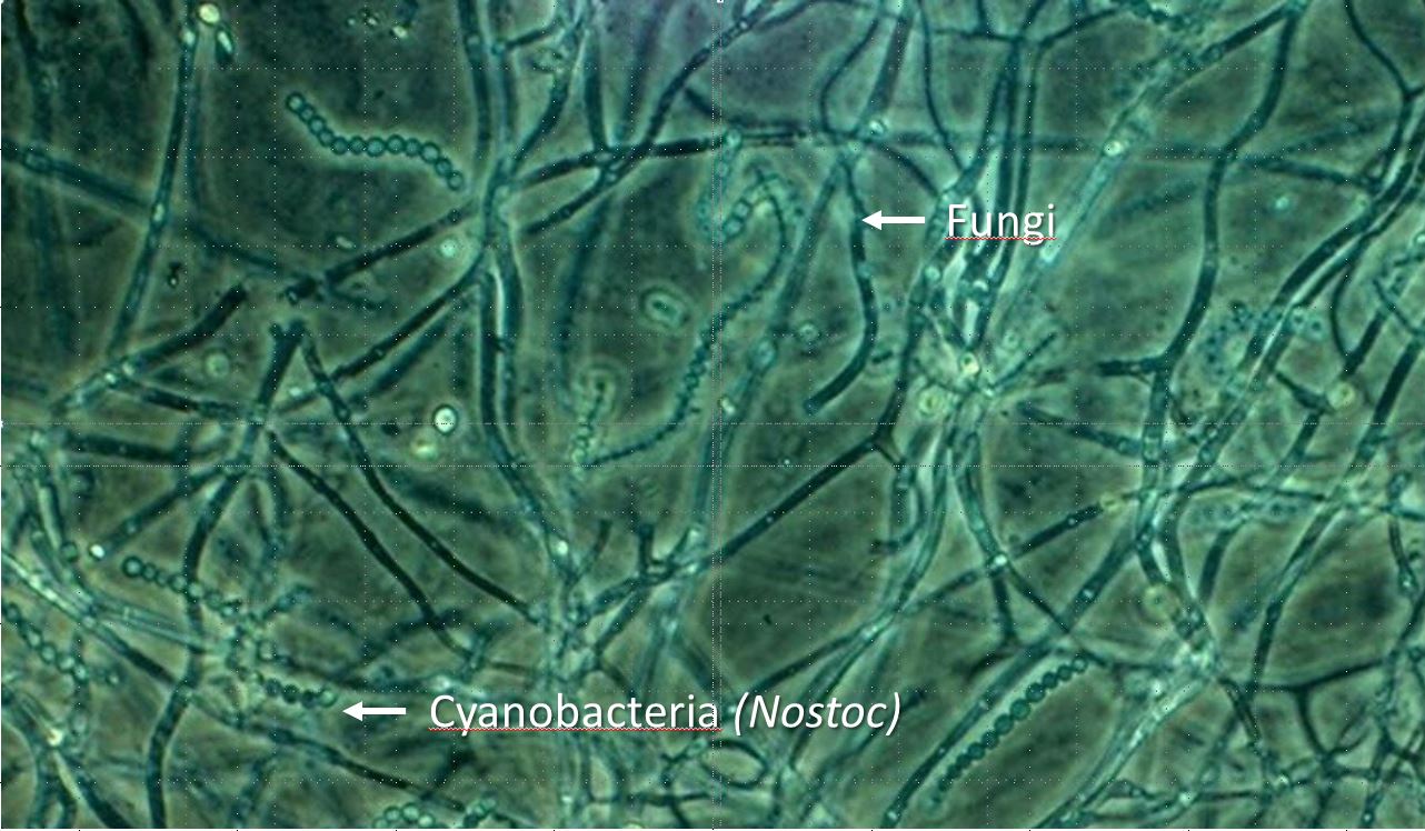
Lichenized fungi belong mainly to the Ascomycetes (Ascomycota). Only a few lichens are formed from Basidiomycetes (Basidiomycota). As for algae, 90% of them are green algae, mainly Trebouxia (50 to 70% of lichens) and Trentepohlia. The rest is represented by cyanobacteria, of which the genus Nostoc is the most common. But this association is much more complex (see Lichens, surprising pioneering organisms).

1. How is a lichen built?

In gelatinous lichens such as Lathagrium, the fungus and cyanobacteria are homogeneously mixed : we speak of a homeomerous structure (Figure 1). In other lichens, the two partners are distributed differently to form a so-called heteromerous structure. In foliose lichens, for example, the structure shows layers (heteromerous stratified structure).

lichens - phaerophyscia orbicularis
Figure 2. Heteromerous structure. Vertical section of foliose thallus of Phaeophyscia orbicularis. [Photo source © Danièle Gonnet]
Figure 1. Homeomerous structure of Enchylium tenax with gelatinous thallus [Source: document © J. Asta]
 

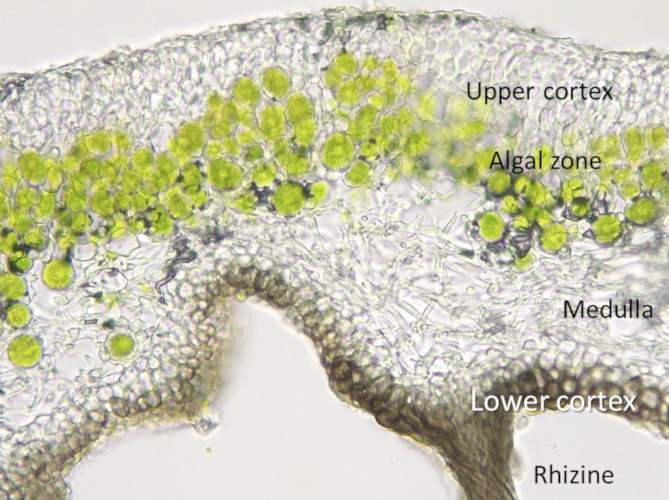
On a vertical section of the thallus, we recognize from top to bottom (Figure 2):

  • an upper cortex formed only by fungi;
  • an algal zone where fugi filaments and algae cells are intertwined;
  • a medulla or medullary layer [1] where there are only fungi filaments;
  • a lower cortex formed only by fungi from which escape filaments or rhizinae used to fix the thallus on the substrate.

2. Birth of a lichen

Two modes of reproduction can take place: vegetative and sexual reproduction.

Xanthoria parietina - lichens
Figure 3. Vegetative propagation: left, isidia from Parmelia saxatilis; right, Lepra amara thallus with soredia [Source photos © J. Asta]
Vegetative propagation is carried out by fragmenting the thallus or by means of natural cuttings. In Lepra amara, for example, the thallus is dotted with floury spots, soralia. At each soralium, the cortex is interrupted and dusts -formed by the fungus filaments and algae- or soredia escape, which can be carried by rain, wind, animals, etc., and thus spread the species. The isidia come from small expansions of the thallus whose cortex persists. When thallus strips rub against each other or insects pass through, these isidia can detach and also be used for dissemination (Figure 3).

Xanthoria parietina - lichens
Figure 4. Example of the development cycle of Xanthoria parietina. 1: adult thallus with apothecium; 2: vertical section of an apothecia, showing the hymenium (h); 3: detail of the hymenium in section (e: epithecium ; p: paraphyses ; s: spores ; a : asci); 4 : mature spores from the hymenium ; 5 : algal cells (Trebouxia) ; 6 : spores in germination ; 7 : primordial stage of the thallus ; 8 : thallus in the process of differentiation. [Source: drawings © J. Asta]
The algae divides only by mitosis (asexual reproduction), only the fungus has sexual reproduction. Let’s take the example of Xanthoria parietina (Figure 4). The reproductive or apothecium organ has a fertile or hymenium tissue consisting of asci where the spores originate. Each asci contains 8 spores with 2 unicellular lodges separated by an equatorial thickening giving the spores an hourglass shape. Between the asks are sterile or paraphyses filaments, the coloured end of which extends beyond the top of the asks and constitutes the epithecium.

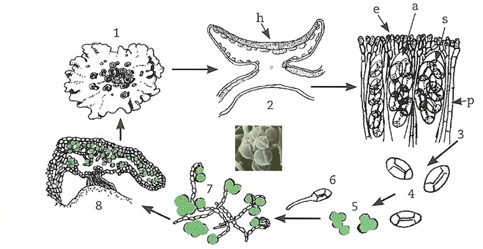
At maturity, the spores are violently expelled from the ascus then germinate on the substrate forming mycelial filaments. For a lichen to recover, the mycelium thus formed must meet a single algae. A new thallus can therefore be rebuilt and give back an adult thallus. The symbiosis then gradually recovers.


Notes and references

Cover image. Xanthoparmelia conspersa. [Source: © J. Asta]

[1] In crustose lichens, the medulla consists of loosely packed hyphae that are inseparable from the substrate.

| Focus /3 | Lichens, surprising pioneering organisms

地衣:共生生物

PDF

  地衣被认为是真菌的“近亲”,传统意义上,它们是由真菌绿藻蓝藻,或与绿藻和蓝藻的结合组成的。这种关系被称为共生,真菌是真菌共生体,藻类是光合共生体。地衣真菌主要属于子囊菌纲(子囊菌门)。只有少数地衣是由担子菌(担子菌门)形成的。藻类中90%为绿藻,主要为共球藻属(50—70%的地衣)和橘色藻属。其余以蓝藻为代表,其中念珠藻属是最常见的。这种共生关系很复杂(参见《地衣,令人惊讶的开拓者》)。

1.地衣是如何形成的?

  在胶状地衣(如胶耳衣)中,真菌和蓝藻是均匀混合的:我们称之为结构(图1)。在其他地衣中,这两个伙伴的分布不同,形成了所谓的异结构。例如,在叶状地衣中,其结构呈层状(异层结构)。

图1. 同层结构。胶状地衣体的同层结构。[图片来源© J. Asta]
图2. 异层结构。圆叶黑蜈蚣衣垂直切面。[图片来源© Danièle Gonnet]

  在地衣体的垂直切面上,我们可以看到(图2,顺序从上到下):

  • 上皮层仅由真菌形成;
  • 藻胞层为真菌菌丝与藻细胞交织区域;
  • 髓层仅有菌丝存在[1]
  • 下皮层仅由真菌形成,从其中逸出用于在基质上固定地衣体的丝或

2.地衣的繁殖

  地衣有两种繁殖方式:无性繁殖和有性繁殖。

图3. 无性繁殖: 左图,藻纹梅花衣的裂芽;右图,带着粉芽的Lepra amara
[图片来源: © J. Asta]

  无性繁殖是通过地衣体的裂片和自然断裂进行的。例如,Lepra amara地衣体上点缀着粉状斑点,即粉芽。每个粉芽堆的皮层裂开,真菌丝和藻类形成的粉尘和粉芽从中逸散,这些粉尘可以被雨、风、动物等携带,从而传播繁殖。皮层仍然存在的地衣体扩张后形成裂芽,当地衣体相互摩擦,或有昆虫通过时,这些裂芽可以分离,也可以传播(图3)。

  藻类只进行有丝分裂(无性生殖),只有真菌进行有性繁殖。此处以丽石黄衣为例(图4)。丽石黄衣的生殖器官或子囊器官有一个可育的子实层组织,由子囊组成,子囊可以产生孢子。每个子囊含有8个孢子,2个单细胞腔,由中间增厚层隔开,使孢子呈沙漏状。在子囊之间是不育的或侧丝碎片,其有颜色的末端延伸出子囊的顶端,构成囊层被。

图4. 丽石黄衣生长过程的图例:1:带子囊盘的成熟地衣体;2:子囊盘的垂直截面,显示子囊层(h);3:子囊层的详细图解(e:囊层被;p:侧丝;s:孢子;a:子囊);4:成熟的孢子来自子囊层;5:藻类细胞(共球藻);6:孢子萌发;7:地衣体幼年时期;8:分化过程中的地衣体。 [图片来源: © J. Asta]

  成熟后,孢子从子囊中猛烈地排出,落在基质上发芽后形成菌丝。为了使地衣恢复,这样形成的菌丝体必须与单个藻类相遇。这样一来,一个新的地衣体可以产生,并长成一个成年的地衣体,这种共生关系便可以逐渐恢复。


注释及引用:

封面图片:黄梅地衣(Xanthoparmelia conspersa)。[来源: © J. Asta]

[1] 壳状地衣的髓层由松散堆积的菌丝组成,与基质密不可分。