蝙蝠和病毒如何和谐共处

  长期以来,蝙蝠一直被人们所忽视甚至蔑视。但近年来,它们引起了公众和科研人员的关注。蝙蝠非凡的飞行及回声定位等能力让公众大感兴趣。由于蝙蝠体型很小,最重要的是,它们独特的免疫系统使它们可以耐受许多陆生哺乳动物难以招架的病毒,因此极其长寿,这使蝙蝠成为了研究人员绝佳的研究模型。然而,蝙蝠与致病性病毒之间的联系却毁了它们的良好形象。的确,蝙蝠似乎近年来在各类新型病毒流行病中充当了关键角色,这些流行病近年来越来越频繁地撼动着世界人口。包括当前正大规模肆虐的COVID-19,再次把蝙蝠推向了风口浪尖。该事件引出了很多疑问:蝙蝠是怎样成为致病性病毒的首选宿主的?它们在COVID-19大流行的起源中扮演了什么角色?人类和家畜越来越多地收到新型病毒性疾病的影响,它们是否真的对此负有责任?

1. 聚光灯下的蝙蝠

环境百科全书-蝙蝠-蝙蝠
图1. 蝙蝠是很多致病新型病毒的天然宿主。
[来源:亨尼巴病毒属,CSIRO, CC BY 3.0,通过维基共享 /丝状病毒科,图片来源:CDC/辛西娅·戈德史密斯(Cynthia Goldsmith),公共领域,通过维基共享 /冠状病毒科,公共领域]
(Henipavirus:亨尼巴病毒属;Filovirus:丝状病毒科;Coronavirus:冠状病毒科;Nipah:尼帕;Hendra:亨德拉;Ebola:埃博拉;SARS:严重急性呼吸综合征;MERS:中东呼吸综合征;COVID-19:2019冠状病毒病)

  2020年初出现一种呼吸道疾病,这种疾病在世界人口范围内迅速传播,这是继2009-2010年的传播范围相对有限的甲型流感之后,人们不得不在21世纪面对的首次大型流行病(见现代的病毒大流行)。这次大型流行病的罪魁祸首也是一种冠状病毒,与2002-2003年在中国造成呼吸道感染流行的病原体相似。该病毒在后来被称为严重急性呼吸道综合征冠状病毒或SARS-CoV。最近,为了区别于新的SARS-CoV-2,SARS-CoV被重新命名为SARS-CoV-1,而SARS-CoV-2就是由世界卫生组织(WHO)命名的在2019年传播的冠状病毒病(Corona Virus Disease 2019)COVID-19的罪魁祸首。

  与SARS-CoV-1相同,蝙蝠似乎也出现在了病毒传播链上。事实上,初步研究表明,SARS-CoV-2被认为是蝙蝠冠状病毒[1]和马来穿山甲(Manis javanica)冠状病毒的重组的结果[2]。后者就如同2002-2003年传染病流行事件的果子狸(Paguma larvata),被认为是SARS-CoV-2在蝙蝠和人类传播链上的中间宿主,导致了病毒在人类中的高度传染性(见焦点蝙蝠和冠状病毒的出现)。

  这不是蝙蝠第一次受到关注。首次从蝙蝠身上分离出病毒要追溯到到1911年。当时人们对狂犬病感兴趣,并且发现了狂犬病是由巴西常见的吸血蝙蝠传播的。而后蝙蝠与病毒的联系便淡出了人们的视野,直到1990-2000年间,健康监测计划再次发现蝙蝠是许多新型病毒天然宿主,如亨尼巴病毒属(亨德拉和尼帕),丝状病毒科(埃博拉)和冠状病毒科(SARS)[3](图1)。出乎意料的是,人们发现蝙蝠不受大部分病原体的影响。研究甚至已经证明,它们可以承受对其他哺乳动物来说致命的病毒载量,尤其是亨德拉和尼帕病毒[4]

2. 蝙蝠与病毒?

2.1. 蝙蝠占哺乳动物总数的四分之一

环境百科全书-蝙蝠-翼手目
图2. 翼手目(蝙蝠)物种占所有现生哺乳动物的近四分之一。
[来源:阿拉纳(Aranae),公共领域,通过维基共享]
(Marsupials:有袋类;Artiodactyla:偶蹄目;Carnivora:食肉目;Primates:灵长目;Insectivores:食虫目;Rodents:啮齿目;Bats:蝙蝠)

  蝙蝠是唯一会飞的哺乳动物,在分类学上归为翼手目,这个名字来自于希腊语kheir,意思是“手”和“翅膀”,即“用手飞行”。该目包含大量物种,目前超过1428多种,约占6495多种已知哺乳动物的四分之一,仅次于啮齿动物(图2)。

  近期基于分子学数据修订了翼手目的分类,将其重新分为了系统发育更连贯的两个亚目(见焦点蝙蝠和冠状病毒的出现):

  • 环境百科全书-蝙蝠-翼手目
    图3. 眼镜狐蝠(Pteropus conspicillatus)于凯恩斯,澳大利亚。它们主要在热带雨林的树冠层中生活。
    [来源:©弗朗索瓦·穆图(François Moutou)]
    阴翼手亚目(狐蝠形类),包括热带的果蝠或是狐蝠(图3)。
  • 阳翼手亚目(蝙蝠形类),包括除五个科外的所有原小蝙蝠亚目的成员。

2.2. 形式各样的生态位

环境百科全书-蝙蝠-飞行中的小菊头蝠
图4. 飞行中的小菊头蝠(Rhinolophus hipposideros)。这种动物喜欢由灌木丛和牧草地构成的树篱。[来源:©路易·玛丽·普雷奥(Louis-Marie Préau)]

  蝙蝠有着异常丰富的物种多样性,分布在除南北极外的所有大陆,并占有形式各样的生态位(图3、4)。在饮食方面,它们多以水果和虫类为主,但也可能吃花蜜、吃花粉、吃肉(鱼、两栖动物、小型哺乳动物)、杂食或以吸血为生。吸血为食的仅限于生活在美洲热带地区的三种蝙蝠。尽管这些吸血蝙蝠很少攻击人类,但却仍然成为了人们恐惧的对象。不过它们可以通过吸食哺乳动物的血液来传播狂犬病病毒。因此,在进一步介绍之前,我们可以知道蝙蝠是一个很有利于病毒繁殖、交换传播的“培养基”。

2.3. 什么是蝙蝠的病毒组?

环境百科全书-蝙蝠-蝙蝠病毒分析
图5. 两种蝙蝠病毒组的分析。[来源:本作作者]
(Salivary virome:唾液病毒组;Faecal virome:粪便病毒组)

       最近对蝙蝠与病毒的重新研究将会激发研究人员对这种从生态和健康角度治疗疾病的新方法的兴趣。此外,前沿的研究手段也将投入这股热潮,使用宏基因组和高通量测序的方式,可以在极短时间内对生物样本的全基因组(病毒组)进行分析,且无需对样本进行培养。这些前沿技术将会使研究计划加速,其目的是提升人们对蝙蝠病毒组的认知(图5)。因此,研究者们将在允许进行蝙蝠研究的国家开展一系列活动,收集各类样本,包括粪便(排泄物)、尿液、口腔或肛拭子,甚至是器官[5]

  通过这些生物样本,大量的病毒序列被识别,其中的一些与已知的人类致病病毒序列相似[6]。但我们不能像有些人那样草率地得出结论,认为蝙蝠是导致人类所有病毒性疾病发生的罪魁祸首!虽然这些研究手段大大提高了我们对病毒与蝙蝠之间联系的认识,但是鉴于分析手段的缺,我们在解释这些数据时必须要保持谨慎。

  然而,令人惊讶的是,最新研究进展证实了1974年的简单观察结果,即蝙蝠对携带各类RNA和DNA病毒的倾向性[7]

2.4. 蝙蝠是“病毒株”吗?

环境百科全书-蝙蝠-蝙蝠体内病毒的多样性
图6. 蝙蝠体内病毒的多样性。数量最多的是冠状病毒,但研究才刚刚开始。
[来源: 列科(Letko)等制作的饼状图[11]版权/链接: doi: 10.1038/s41579-020-0394-z]
(Filoviridae:丝状病毒科;Parvoviridae:细小病毒科;Picornaviridae:微小核糖核酸病毒科;Flaviviridae:黄病毒科;Herpesviridae:疱疹病毒科;Circoviridae:圆环病毒科;Reoviridae:呼肠孤病毒科;Polyomaviridae:多瘤病毒科;Adenoviridae:腺病毒科;Astroviridae:星状病毒科;Rhabdoviridae:炮弹病毒科;Hepadnaviridae:肝病毒科;Papillomaviridae:乳头瘤病毒科;Hantaviridae:汉坦病毒科;Coronaviridae:冠状病毒科;Paramyxoviridae:副黏液病毒科;Virus families with fewer than 50 different sequences:差异序列数目少于50的病毒科:Caliciviridae:杯状病毒科;Peribunyaviridae:本雅病毒科;Nairovridae:内罗病毒科;Unclassified viruses:未分类病毒;Retroviridae:逆转录病毒科;Hepeviridae:肝炎病毒科;Orthomyxoviridae:正黏液病毒科;Phenuiviridae:白细病毒科;Poxviridae:痘病毒科;Pivobirnaviridae:皮可比那病毒科;Togaviridae:披盖病毒科;Genomoviridae:类双生病毒科;Bornaviridae:玻那病毒科;Anelloviridae:指环病毒科;Unclassified ssDNA viruses:未分类的单链DNA病毒;Unclassified Bunyavirales:未分类的本雅病毒目)

  虽然啮齿动物就数量而言是哺乳动物中的第一大目,但一项关于啮齿类动物和翼手目动物中存在的人畜共患病毒数量的比较研究[8]表明,啮齿动物中的人畜共患病毒更多,为68(啮齿目)比61(翼手目),但每个物种的病毒总数使翼手目动物位居榜首,即1.79(翼手目)比1.48(啮齿目)。最近,彼得·达扎克(Peter Daszak)等在自然Nature[9]上发表的研究表明,可用感染哺乳动物的病毒总数来预测引起人畜共患病病毒的比例。而这项研究的一个结论就是:相比于哺乳纲其他目的动物,蝙蝠所携带的人畜共患病毒的比例更高

  特别是在冠状病毒方面,另一项大型比较研究[10]是在19000多只包括蝙蝠,啮齿动物与非人的灵长类动物中进行的,研究结果表明98%的冠状病毒来自于蝙蝠。在已经发现的100种冠状病毒中,就有91种来自于翼手目,可见冠状病毒的数量之大、种类之多。在呈冠状病毒阳性的个体中,蝙蝠占8.6%,而在其他动物中仅占0.2%。从这些结果中我们可以推断出全球范围内的所有种的蝙蝠至少携带了3200种冠状病毒。最近重新评估的数字为3796种(图6)[11]。蝙蝠似乎具有特殊的能力。

  不过,由于研究中存在抽样偏差,所以各种情况都应加以考虑。作为哺乳纲中排行第二的,拥有1428个物种的翼手目,蝙蝠不可避免地位居前列。最近,研究人员调查了哺乳动物和鸟类,他们发现,不论宿主是属于鸟纲还是哺乳纲,病毒性人畜共患病的风险都是相同的,所以每个目患人畜共患病的数目随物种基数的增加而增加[12]。因此,对于蝙蝠和啮齿动物来说,人畜共患病毒大量存在仅仅与它们庞大的物种基数有关。

  事实上,蝙蝠在病毒出现的过程中扮演的特殊角色更可能从它们的特殊生态行为、不同寻常的生理机制与免疫特征以及它们强于其他动物的RNA病毒交换能力中寻找答案。最后,我们还必须补充的一点,人类通过在生态系统中的行为给这些因素的协同作用创造了有利条件

3. 为什么蝙蝠是致病性病毒们的首选宿主?

3.1. 蝙蝠和病毒已经共存很久了

环境百科全书-蝙蝠-古老的蝙蝠
图7. 古老的蝙蝠。A.芬尼氏爪蝠(Onychonycteris finneyi)化石,最古老的翼手目化石之一(皇家安大略博物馆),可追溯到5250万年前(始新世)。发现于北美的怀俄明州。这种蝙蝠会飞也会爬树。这种蝙蝠还没有回声定位能力,因此使用视觉和嗅觉捕食昆虫。
[来源:A, 马修·迪利翁(Matthew Dillon), CC BY 2.0, 通过网络相簿]。B.芬尼氏爪蝠的复原图。[© N.田村(N. Tamura); CC BY-SA许可证]

  蝙蝠/病毒的共生关系可能由来已久,因为最古老的蝙蝠化石出现于5000万年前的始新世(图7)。尽管白垩纪-第三纪物种大灭绝仅仅发生1000万年前,但始新世的蝙蝠的多样性已经十分显著。蝙蝠的起源一直是个谜题,直到最近的一篇文章[13]证实了翼手目的古老程度,并阐明了蝙蝠的起源。通过比较六种蝙蝠的基因组和其他种类的哺乳动物基因组,研究人员发现翼手目属于劳亚兽总目(有胎盘类哺乳动物的一个总目),翼手目与猛兽有蹄超目这个演化支有共同祖先,猛兽有蹄超目包括骆驼类、猪类、鲸类、马类、犀类、食肉类和穿山甲类,从共同祖先处一共分出了五个目(翼手目、食肉目、鳞甲目、奇蹄目、偶蹄目),这将翼手目出现的时间推到了6500多万年前

3.2. 病毒和蝙蝠的长期共同演化

  因此,病毒有一个漫长的时期来适应它们的蝙蝠宿主并与其共同演化。病毒感染宿主的能力经受长期的自然选择,致使其对存在于蝙蝠细胞表面特定分子的适应性越来越强。这些表面分子俗称受体,它们在病毒侵入宿主细胞的过程中起重要作用。此外,由于这些受体分子参与了许多基础的生理过程,导致它们在动物的世界里一直有很高的保守性。这就解释了为何病毒能轻易能轻易跨越物种障碍,并迅速在哺乳动物宿主中传播。

环境百科全书-蝙蝠-SARS-CoV-2
图8. SARS-CoV-2的刺突蛋白与宿主细胞受体(由血管紧张素I转换酶2(ACE2)组成)结合,宿主细胞膜蛋白酶将刺突蛋白裂解激活后,病毒复合物直接在细胞表面发生融合(A)。无膜蛋白酶,则病毒通过胞吞进入宿主细胞(B)。[来源:本作作者]
(Spike protein:刺突蛋白;RBD site:受体结合域;ACE2 Receptor:血管紧张素I转换酶2受体;Protease:蛋白酶;Menbrane fusion:膜融合;Endocytosis:胞吞;Host cell:宿主细胞;SARS-CoV-2:严重急性呼吸综合征冠状病毒2)

  以SARS-CoV-2为例,受体很快被识别为血管紧张素I转换酶2(ACE2)(图8)。ACE2参与血压调节,广布于脊椎动物体内以及许多组织和器官(肺、心脏、动脉、肾脏以及消化系统)的细胞表面。在感染过程中,病毒蛋白S(Spike意为刺状的)作为钥匙与ACE2这把锁相互作用。这些刺状物给予了病毒无上的冕冠,因此这种病毒被称为冠状病毒。

  然而,这把钥匙(S蛋白)必须在其受体结合域(RBD)上持续变异,从而获得与人类ACE2受体相同的特定氨基酸序列和一个可被宿主酶切的特定位点,这样SARS-CoV-2才能感染人类的细胞[14]

  这些突变可能是蝙蝠冠状病毒寄宿在中间宿主时发生的,最终形成了SARS-CoV-2。除了人类,冠状病毒也有能力感染其他哺乳动物,例如灵长类(猕猴)、某些种类的鹿和鲸、家猫、老虎、黄金仓鼠、雪貂、水鼬以及家犬。除了哺乳纲的动物,脊椎动物亚门中其他纲的动物似乎不会被冠状病毒感染[15]

3.3. 飞行对蝙蝠和病毒间的联系至关重要

环境百科全书-蝙蝠-飞行中的马铁菊头蝠
图9. 飞行中的马铁菊头蝠(Rhinolophus ferrumequinum)。
[来源:©路易·玛丽·普雷奥(Louis-Marie Préau)]

  在翼手目众多适应性状的演化中,飞行能力的获得最为引人注目(图9)。飞行似乎在蝙蝠和病毒的联系中起了重要作用。这种能力不仅仅是蝙蝠绝佳的运动方式,它同样使病毒得以在大范围区域内和不同的生态系统中进行传播。例如,研究发现,亨尼巴病毒属病毒的地理分布区域与狐蝠属蝙蝠的活动区密切重叠,而关于人和动物的病例在澳大利亚、孟加拉国、印度、马来西亚和新加坡均有报导。

  更令人惊讶的是,飞行极大地促进了蝙蝠的新陈代谢(相比于鸟类的2次,蝙蝠可达15-16次)以及提高了蝙蝠的体温(>38℃),使蝙蝠近乎时刻处于发烧状态。但是,发烧是内温动物用于抑制病原体增殖、刺激免疫系统工作的天然防御机制。因此,通过诱导或多或少的长期发烧状态,飞行有助于提高蝙蝠对病毒的抵抗力[16]

3.4. 呼吸与氧化应激

环境百科全书-蝙蝠- 活性氧
图10. 活性氧(ROS)在线粒体内膜上的电子传递链中产生,它可以诱发氧化应激使膜脂、蛋白质和DNA在细胞层次上发生改变。细胞有抗氧化剂和修复系统来进行自我保护。[来源:本作作者]
(Respiratory chain:电子传递链;Mitochondrial DNA:线粒体DNA;Mitochondria:线粒体;Nuclei:细胞核;Nuclear DNA:核DNA;Cell:细胞;Implementing antioxidant systems:抗氧化系统的运作;Oxydative stress:氧化应激;Mutations and deletions in mitochondrial and nuclear DNA:线粒体DNA与核DNA的突变与缺失;Implementing repair systems:修复系统的运作;Membrane alteration:膜改造)

  在飞行过程中,蝙蝠消耗的氧气是正常情况下的四倍,从而在电子传递链中产生大量的活性氧(ROS),导致剧烈氧化应激的产生(图10)。氧化应激通常会对包括DNA在内的细胞成分造成严重损伤,但这对蝙蝠似乎影响甚微。事实上,蝙蝠已经选择了十分高效的线粒体抗氧化系统来维持自身飞行的高能量需求,并可以在氧化应激中保护自己[17]。当损伤出现时,蝙蝠可依赖自身的高性能DNA修复系统[18]。由于许多病原体在感染初期就会使机体出现氧化应激,所以线粒体在免疫系统中的作用愈发被认可,这些应性状是在进化过程中慢慢被蝙蝠选择,对翼手目的免疫抑制病原体产生了有益的影响[19]

3.5. 蝙蝠的长寿有利于病毒长期存在

环境百科全书-蝙蝠-779种哺乳动物寿命系数
图11. 779种哺乳动物寿命系数(寿命观测值与寿命理想值之比)与体重(以克为单位)的函数关系。黑色虚线代表系数为1。大多数蝙蝠的寿命与其体重有关(蓝点),就像一种啮齿动物裸鼹鼠(黑星)一样。对几种蝙蝠(黑色剪影)的端粒长度与其寿命的关系进行了估算。
[来自弗利(Foley)等[20], CC BY – NC 4.0 / 链接: https://advances.sciencemag.org/content/4/2/eaao0926]
(Longevity quotient:寿命系数;Log body mass:体重量;Bats:蝙蝠;All other mammals:其他哺乳动物;Naked mole rat:裸鼹鼠;Bats-telomeres measured:蝙蝠端粒的检测;Myotis myotis:大鼠耳蝠;Rhinolophus ferrumequinum:马铁菊头蝠;Myotis bechsteinii:长耳鼠耳蝠;Miniopterus schreibersii:长翼蝠)

  与此同时,病毒会逐渐和其蝙蝠宿主相适应,导致病毒自身的致病力降低。换句话说就是双方都学会了如何和谐共处。由此可见,蝙蝠的许多生命特征都有利于病毒长期存在

  我们在上文中已经知道了蝙蝠可以调节氧化应激,而氧化应激常常会导致慢性炎症,加速衰老。同样,端粒(染色体终端)的缩短常常导致衰老和癌症的出现,但我们仍未在蝙蝠身上观察到这些现象[20]。因此,蝙蝠有着悠长的寿命(图11),几乎没有衰老的迹象,患癌的概率也微乎其微。在19种寿命超过人类的哺乳动物中(考虑体型因素),有18种是蝙蝠,而体重7克寿命却超过41岁的布氏鼠耳蝠(Myotis brandtii)是其中的佼佼者。蝙蝠的这种长寿能力有利于病毒在其个体中持续存活,也有利于病毒长期在幼体和成体之间进行交换。

环境百科全书-蝙蝠-冬眠中的达马拉菊头蝠
图12. 冬眠中的达马拉菊头蝠。A.达马拉菊头蝠群体;B.达马拉菊头蝠(Rhinolophus ferrumequinum)在翅膀的包裹下昏睡(我们可以看到它的鼻叶,鼻叶可以使鼻孔发出的超声波集中,以此在运动时进行定位)。
[来源:©马克·皮赫特(Marc Pihet)]

3.6. 睡眠与迟滞状态

  另一个有利于病毒在蝙蝠中长存的因素是蝙蝠的睡眠迟滞状态(图12、13)。

  睡眠是温带国家的蝙蝠在寒冷季节食物短缺的情况下选择的生存策略,如鸟类的其他动物则选择向温暖地区迁徙。

环境百科全书-蝙蝠-睡眠中的伊氏菊头蝠
图13. 睡眠中的伊氏菊头蝠(Rhinolophus hipposideros),它用爪子将身体悬挂在墙上。
[来源:©马克·皮赫特(Marc Pihet)]

  因此,在为时数月的冬眠期间,蝙蝠会进入一种体温极低、新陈代谢极慢的深度睡眠状态,以此保存能量。免疫防御功能也随之减弱;这种变化有利于病毒在蝙蝠体内生存。这样就能解释病毒传播的季节性了。至于那些生活在热带、不进行冬眠的蝙蝠,据观察,它们会周期性地陷入白天迟滞状态,同时新陈代谢降低,就像冬眠时一样[17]

3.7. 共生的重要性

  病毒的持久存在可以表现在个体层面和群体层面上。翼手目的动物有强烈的集群生活倾向性,它们可以成百甚至上千只不同年龄、有时是不同种的个体组成集群。目前已知的最大的哺乳动物聚集地在德克萨斯州的布莱肯洞穴,那里聚集了两千多万只巴西犬吻蝠(Tadarida brasiliensis mexicana)(图14)。洞穴里的蝙蝠需要至少三个小时才能全都出来!

环境百科全书-蝙蝠-巴西犬吻蝠
图14. A,布莱肯洞穴(德克萨斯州,美国)是全世界规模最大的蝙蝠聚集地之一,每年3-10月约有两千多万只巴西犬吻蝠在此聚集。B,巴西犬吻蝠(Tadarida brasiliensis)
[来源:A,摄影:乔纳森·阿隆佐(Jonathan-Alonzo),该照片由国际蝙蝠保护协会提供/ B,摄影:迈克尔·达勒姆·明登(Michael Durham Minden),该照片由国际蝙蝠保护协会提供]。

  显然,蝙蝠的这种聚集现象使得病毒可以轻而易举地进行传播,要么通过蝙蝠互舔毛发(它们的毛发常常被尿液、粪便弄脏),要么通过体液(尿液、唾液)的气溶胶化,这是病毒传播的良好载体。为了在夜间活动和进行捕猎,一些蝙蝠会使用回声定位(见焦点回声定位),这种方法可以调动耳朵、鼻子和喉咙,并从鼻粘膜分泌物和唾液中产生气溶胶,而这都是病毒的首选附着位置。这种在进化中获得的能力包括发射超声波来获得声音的“图像”。

  这些蝙蝠群体证明群体的特点和规模在病毒的维持和变体的出现中扮演了重要的角色。人们普遍认为,蝙蝠成年体和幼体之间的病毒交叉可以促进慢性感染,而不同种个体间的交叉感染可增加病毒的多样性。研究者想知道,这种共生习性是否是导致蝙蝠在耐受病毒的同时强化其独特免疫能力的决定性因素,而这些免疫能力的方方面面直到最近才被发现。

3.8. 独特的免疫能力

环境百科全书-蝙蝠-独特免疫能力
图15. 蝙蝠控制病毒和炎症的独特免疫能力源自飞行。[来源:本作作者]
(Impact of flight on evolution:演化出的飞行能力的影响;Flight:飞行;Metabolism:新陈代谢;Reactive oxygen species:活性氧的种类;Cell damages: DNA, lipids, proteins:细胞损伤:DNA,脂质,蛋白质;Positive selection:正向选择;Reduction of inflammation in bats:蝙蝠炎症的减少;Mobilisation of interleukins to curb the interferon response:动员白细胞介素来抑制干扰素响应;decrease in the interferon regulation factor(IRF3):干扰素调节因子的降低;Decrease in the interferon beta:干扰素β的降低;Low level of inflammation:炎症等级降低;Advantageous for virus replication:利于病毒复制;Bat cell:蝙蝠细胞;Virus replication:病毒复制;Virus:病毒;Control of virus attack:抵御病毒攻击;Higher basal level for interferon expression in bats:蝙蝠干扰素更高基础等级的表达)

  一般情况下,人类和大部分哺乳动物都在感染病毒时启动免疫系统来响应病毒的攻击。但蝙蝠并非如此,它们能在低响应水平永久维持这种免疫状态,因此在感染发生时它们可以迅速变得强壮起来(图15)。在最初的炎症反应中,病原体到达宿主细胞导致细胞因子(干扰素)的靶向发射,随之而来的第二步反应是使用白细胞介素缓解炎症,以防止病原体对宿主产生不利影响。事实上,人们认为正是飞行这种剧烈的促炎症活动诱发了蝙蝠获得这种防止炎症加剧的独特的免疫能力[18]。细胞因子在新冠病毒流行中曾导致患者出现免疫风暴,致使一些病患体内出现严重的炎症反应,急救人员试图像蝙蝠一样去控制这种现象,因为这种严重的炎症反应会严重威胁患者生命[21]

  事实上,蝙蝠似乎有能力抑制与它们长期共同进化病原体的免疫反应,以限制感染后的免疫病理影响[22]。蝙蝠和病毒似乎签订了互不侵犯条约。

  最近对六个蝙蝠的基因组[13]的分析显示了多种内源性病毒,这个结果似乎可以证实蝙蝠病毒的耐受性,可以认定病毒是蝙蝠自身的组成部分。此研究还揭示了炎症反应集中在数量有限的基因上,并且确认了与病毒感染的耐受性相关的基因的存在。这项研究旨在让我们更好地理解蝙蝠是怎样耐受冠状病毒感染。

4. 蝙蝠该为病毒爆发负责吗?

  对蝙蝠在病毒流行中的作用的认识可能使人们将病毒爆发的责任推给它们,并会考虑通过消灭蝙蝠来解决问题。然而这种直观的反应很不合适,甚至不利于人类的健康。这一点在乌干达已经得到了验证。为了预防马尔堡病毒的感染,人们消灭了生活在矿井中的果蝠。但是,其他具有易感性的蝙蝠重新占据了这些地方,并且将更多样化的病毒重新引入到这些新种群中。

  这样的反应全然忘记了翼手目的蝙蝠也是生态系统功能中不可缺失的一环。在热带地区,它们扮演着给植物授粉和远距离传播种子的角色。人们利用蝙蝠,并把蝙蝠的粪便作为肥料。而食虫蝙蝠在调节昆虫数量方面起着重要作用,从而减少杀虫剂的使用。蝙蝠对环境变化十分敏感,所以也是极好的环境健康指示器

环境百科全书-蝙蝠-人畜共患病的有利因素
图16. 人畜共患病的有利因素。[来源:本作作者]
(Extrinsic factors:外部因素;drought:干旱;habitat loss:失去栖息地;bush fires:丛林火灾;cave destruction:洞穴破坏;Intrinsic factors:内部因素;age:年龄;pregnancy:妊娠;nutritional status:营养情况;Urbanization:城市化;Cultural practices:文化习俗;Eco-tourism:生态旅游;International travel:国际旅行;Trade:贸易;Socioeconomic factors:社会经济因素;Agricultural practices:农业习俗;Bat:蝙蝠;Human:人类;Intermediate host:中间宿主)

       疫情的起因是人类活动对自然生态系统破坏:农业活动的加剧导致滥砍滥伐和生境破碎化、退化和加剧城市化[23](图16)。这些活动侵占了蝙蝠长期生活的天然环境,致使蝙蝠种群更加接近人类栖息地和家畜养殖场[24]。此外,市场上对活体野生或家养动物的售卖使得它们不自然地聚集在恶劣环境中,这是病毒的温床。为了避免这种情况,简单有效的措施是对活体动物市场严加管控。而从长远来讲,应该鼓励人类深刻改变饮食习惯

  以更全面的角度而言,有必要对人类和野生动物及蝙蝠接触的风险因素采取行动。这是新冠病毒大流行给我们的警示。控制这些风险因素需要人类健康、动物健康和生态系统健康领域的紧密合作。换句话说:医生、药剂师、兽医和生态学家,以及经济学家和法学家们必须通力合作来促进生态健康,这是预防未来流行病出现以及防止流行病恶化为健康危机的根本解决途径(见现代的病毒大流行)。

5. 结论

  • 蝙蝠是许多新出现的病毒的天然宿主,但却不受大多数病毒影响。
  • 因为蝙蝠的物种多样性、哺乳动物第二大目、食谱和生态位,它们是病毒的首选宿主
  • 对蝙蝠病毒组(一组病毒的基因组)的研究表明它们对人类病毒性疾病的出现无直接责任
  • 蝙蝠的起源可追溯到6500多万年前,这给予了它们足够的时间和病毒进行共同演化
  • 翼手目的成功基于它们独特的适应性状:飞行和回声定位能力、超常的寿命、嗜睡与迟缓、群居本能和独特的免疫系统,这些都利于它们对感染产生广泛的耐受力
  • 翼手目是生态系统功能中不可缺失的一环,也是良好的环境健康指示器
  • 疫情爆发的原因是人类无节制的活动对自然环境造成了破坏。
  • 为了防止新疾病的流行,必须对人类与野生动物接触的风险因素采取行动。


  感谢弗朗索瓦·穆图(François Moutou)对本文及图片的校对,感谢马克·皮赫特(Marc Pihet)和路易丝-玛丽·普雷奥(Louis-Marie Préau)(www.louismariepreau.com)为本文提供图片说明。


参考资料及说明

封面图片:飞行中的大鼠耳蝠(Myotis myotis) [来源:©路易丝-玛丽·普雷奥(Louis-Marie Préau), www.louismariepreau.com]

[1] Zhou P, Yang XL, Wang XG, Hu B, Zhang L, Zhang W, Si HR, Zhu Y, Li B, Huang CL, Chen HD, Chen J, Luo Y, Guo H, Jiang RD, Liu MQ, Chen Y, Shen XR, Wang X, Zheng XS, Zhao K, Chen QJ, Deng F, Liu LL, Yan B, Zhan FX, Wang YY, Xiao GF & Shi ZL. (2020). A pneumonia outbreak associated with a new coronavirus of probable bat origin. Nature, 579 (7798), 270-273. <doi: 10.1038/s41586-020-2012-7>.

[2] Xiao K, Zhai J, Feng Y, Zhou N, Zhang X, Zou J-J, Li N, Guo Y, Li X, Shen X, Zhang Z, Shu F, Huang W, Li Y, Zhang Z, Chen R-A, Wu Y-J, Peng S-M, Huang M, Xie W-J, Cai Q-H, Hou F-H, Chen W, Xiao L & Shen Y. (2020). Isolation of SARS-CoV-2-related coronavirus from Malayan pangolins. Nature, 583 (7815), 286-289. <doi: 10.1038/s41586-020-2313-x>.

[3] Calisher CH, Childs JE, Field HE, Holmes KV & Schountz T. (2006). Bats: important reservoir hosts of emerging viruses. Clin. Microbiol. Rev., 19 (3), 531-545. <doi:10.1128/CMR.00017-06>.

[4] Middleton DJ, Morrissy CJ, van der Heide BM, Russell GM, Braun MA, Westbury HA, Halpin K & Daniels PW. (2007). Experimental Nipah virus infection in pteropid bats (Pteropus poliocephalus). J. Comp. Pathol., 136 (4), 266-272. <doi: 10.1016/j.jcpa.2007.03.002>.

[5] Hu D, Zhu C, Wang Y, Ai L, Yang L, Ye F, Ding C, Chen J, He B, Zhu J, Qian H, Xu W, Feng Y, Tan W & Wang C. (2017). Virome analysis for identification of novel mammalian viruses in bats from Southeast China. Sci. Rep. 7 (1), 10917. <doi: 10.1038/s41598-017-11384-w>.

[6] Drexler JF, Corman VM, Müller MA, Maganga GD, Vallo P, Binger T, Gloza-Rausch F, Rasche A, Yordanov S, Seebens A, Oppong S, Adu Sarkodie Y, Pongombo C, Lukashev AN, Schmidt-Chanasit J, Stöcker A, Carneiro AJ, Erbar S, Maisner A, Fronhoffs F, Buettner R, Kalko EK, Kruppa T, Franke CR, Kallies R, Yandoko ER, Herrler G, Reusken C, Hassanin A, Krüger DH, Matthee S, Ulrich RG, Leroy EM & Drosten C. (2012). Bats host major mammalian paramyxoviruses. Nat. Commun., 3, 796. <Law: 10.1038/ncomms1796>.

[7] Sulkin SE & Allen R. (1974). Virology. Vol. 8, Basel: Melnick JL, Karger.

[8] Luis AD, Hayman DT, O’Shea TJ, Cryan PM, Gilbert AT, Pulliam JR, Mills JN, Timonin ME, Willis CK, Cunningham AA, Fooks AR, Rupprecht CE, Wood JL & Webb CT. (2013). A comparison of bats and rodents as reservoirs of zoonotic viruses: are bats special? Proc. Biol. Sci. 280 (1756), 20122753. <Law: 10.1098/rspb.2012.2753>.

[9] Olival KJ, Hosseini PR, Zambrana-Torrelio C, Ross N, Bogich TL & Daszak P. (2017). Host and viral traits predict zoonotic spillover from mammals. Nature, 546 (7660), 646-650. <doi: 10.1038/nature22975>.

[10] Anthony SJ, Johnson CK, Greig DJ, Kramer S, Che X, Wells H, Hicks AL, Joly DO, Wolfe ND, Daszak P, Karesh W, Lipkin WI, Morse SS; PREDICT Consortium, Mazet JAK & Goldstein T. (2017), Global patterns in coronavirus diversity. Virus Evol. 3 (1), vex012. <Law: 10.1093/ve/vex012>.

[11] Letko M, Seifert SN, Olival KJ, Plowright RK & Munster VJ. (2020). Bat-borne virus diversity, spillover and emergence. Nat. Rev. Microbiol. 18 (8), 461-471. <doi: 10.1038/s41579-020-0394-z.>.

[12] Mollentze N & Streicker DG. (2020). Viral Zoonotic Risk Is Homogenous Among Taxonomic Orders of Mammalian and Avian Reservoir Hosts. Proc. Natl. Acad. Sci. USA, 117 (17), 9423-9430. <doi: 10.1073/pnas.1919176117>.

[13] Jebb D, Huang Z, Pippel M, Hughes GM, Lavrichenko K, Devanna P, Winkler S, Jermiin LS, Skirmuntt EC, Katzourakis A, Burkitt-Gray L, Ray DA, Sullivan KAM, Roscito JG, Kirilenko BG, Dávalos LM, Corthals AP, Power ML, Jones G, Ransome RD, Dechmann DKN, Locatelli AG, Puechmaille SJ, Fedrigo O, Jarvis ED, Hiller M , Vernes SC, Myers EW & Teeling EC. (2020). Six reference-quality genomes reveal evolution of bat adaptations. Nature, 583, 579-584. <doi: 10.1038/s41586-020-2486-3>.

[14] Zhang T, Wu Q & Zhang Z. (2020). Probable Pangolin Origin of SARS-CoV 2 Associated with the COVID-19 Outbreak. Curr. Biol. 30 (7), 1346-1351.e2. <doi: 10.1016/j.cub.2020.03.022>.

[15] Damas J, Hughes GM, Keough KC, Painter CA, Persky NS, Corbo M, Hiller M, Koepfli KP, Pfenning AR, Zhao H, Genereux DP, Swofford R, Pollard KS, Ryder OA, Nweeia MT, Lindblad-Toh K, Teeling EC, Karlsson EK & Lewin HA. (2020). Broad host range of SARS-CoV-2 predicted by comparative and structural analysis of ACE2 in vertebrates. Proc. Natl. Acad. Sci U.S.A. 2020 Aug 21:202010146. <doi: 10.1073/pnas.2010146117>.

[16] O’Shea TJ, Cryan PM, Cunningham AA, Fooks AR, Hayman DTS, Luis AD, Peel AJ, Plowright RK & Wood JLN. (2014). Bat flight and zoonotic viruses. Emerg. Infect. Dis. 20 (5): 741-745. <doi:10.3201/eid2005.130539>.

[17] Brook CE & Dobson AP. (2015). Bats as special reservoirs for emerging zoonotic pathogens. Trends Microbiol. 23 (3), 172-180. <doi: 10.1016/j.tim.2014.12.004>.

[18] Zhang G, Cowled C, Shi Z, Huang Z, Bishop-Lilly KA, Fang X, Wynne JW, Xiong Z, Baker ML, Zhao W, Tachedjian M, Zhu Y, Zhou P, Jiang X, Ng J, Yang L, Wu L Xiao J, Feng Y, Chen Y, Sun X, Zhang Y, Marsh GA, Crameri G, Broder CC, Frey KG, Wang L-F & Wang J. (2013). Comparative analysis of bat genomes provides insight into the evolution of flight and immunity. Science, 339 (6118), 456-460. <doi: 10 .1126/science.1230835>.

[19] Banerjee A, Baker ML, Kulcsar K & Misra V, Plowright R, Mossman K. (2020). Novel insights into immune systems of bats. Front. Immunol. 11, 26. <doi: 10 .3389/fimmu.2020.00026>.

[20] Foley NM, Hughes GM, Huang Z, Clarke M, Jebb D, Whelan CV, Petit EJ, Touzalin F, Farcy O, Jones G, Ransome RD, Kacprzyk J, O’Connell MJ, Kerth G, Rebelo H, Rodrigues L, Puechmaille SJ & Teeling EC. (2018). Growing old, yet staying young: The role of telomeres in bats’ exceptional longevity. Sci. Adv. 4 (2), eaao0926. <10.1126/sciadv.aao0926>.

[21] Kacprzyk J, Hughes GM, Palsson-McDermott EM, Quinn SR, Puechmaille SJ, O’Neill LAJ & Teeling EC. (2017). A potent anti-inflammatory response in bat macrophages may be linked to extended longevity and viral tolerance. Acta Chiropter, 19 (2), 219-228. <doi: 10.3161/15081109ACC2017.19.2.001>.

[22] Mandle JN, Schneider C, Schneider DS, Baker ML. (2018). Going to bat(s) for studies of disease tolerance. Front Immunol, 9 (2112). < doi: 10.3389/fimmu.2018.02112>.

[23] Gibb R, Redding DW, Chin KQ, Donnelly CA, Blackburn TM, Newbold T & Jones KE. (2020). Zoonotic host diversity increases in human-dominated ecosystems. Nature, 584 (7821), 398-402. <doi: 10.1038/s41586-020-2562-8>.

[24] Afelt A, Frutos R & Devaux C. (2018). Bats, Coronaviruses, and Deforestation: Toward the Emergence of Novel Infectious Diseases? Front. Microbiol. 9, 702. <doi: 10.3389/fmicb.2018.00702>.


环境百科全书由环境和能源百科全书协会出版 (www.a3e.fr),该协会与格勒诺布尔阿尔卑斯大学和格勒诺布尔INP有合同关系,并由法国科学院赞助。

引用这篇文章: LARCHER Gérald (2024年3月13日), 蝙蝠和病毒如何和谐共处, 环境百科全书,咨询于 2024年7月27日 [在线ISSN 2555-0950]网址: https://www.encyclopedie-environnement.org/zh/vivant-zh/bats-viruses-how-live-together-harmony/.

环境百科全书中的文章是根据知识共享BY-NC-SA许可条款提供的,该许可授权复制的条件是:引用来源,不作商业使用,共享相同的初始条件,并且在每次重复使用或分发时复制知识共享BY-NC-SA许可声明。

Bats and viruses or how to live together in harmony

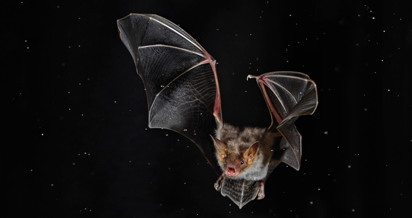
Bats have long been ignored or even despised, and in recent years they have been the subject of increasing public and research attention. Endowed with exceptional abilities such as flight and echolocation, bats intrigue the former. Their unexpected longevity given their size and, above all, a unique immune system that enables them to tolerate many viruses that are poorly tolerated by terrestrial mammals, make them excellent study models for the latter. However, this improved image is tarnished by the knowledge of their links with pathogenic viruses. Indeed, bats seem to play a crucial role in the emerging virus epidemics that have been shaking the world population with increasing frequency in recent years. The current COVID-19 pandemic is no exception, and has once again brought bats to the forefront. This raises many questions: How are bats preferred hosts for pathogenic viruses? What role did they play in the origin of the COVID-19 pandemic? Are they indeed responsible for the emergence of the new viral diseases that are increasingly affecting humans and domestic animals?

1. Bats in the spotlight

Figure 1. Bats are the natural hosts of many emerging viruses responsible for epidemics. [Sources: Henipavirus, CSIRO, CC BY 3.0, via Wikimedia Commons / Filovirus, Photo Credit : CDC/ Cynthia Goldsmith, Public domain, via Wikimedia Commons / Coronavirus, public domain]
The beginning of 2020 was marked by the emergence of a respiratory disease that rapidly spread among the world’s population to such an extent that it had to be recognized that humans were facing, after the relatively limited influenza A pandemic in 2009-2010, the first major pandemic of the 21st century (See Viral Pandemics of the Modern Era). It turns out that the pathogen responsible for this pandemic is a coronavirus, similar to the one that caused an epidemic of respiratory infections mainly localized in China in 2002-2003. The latter was then called severe acute respiratory syndrome coronavirus or SARS-CoV. It has recently been renamed SARS-CoV-1 to differentiate it from the new SARS-CoV-2, which is responsible for the disease named by the World Health Organization (WHO) COVID-19 for COronaVIrus Disease 2019.

As with SARS-CoV-1, bats appear to be involved in the viral transmission chain. Indeed, preliminary results have shown that SARS-CoV-2 is thought to result from the recombination of a bat coronavirus [1] with a Malayan pangolin (Manis javanica) coronavirus [2]. The latter, like the masked palmed civet (Paguma larvata) in the 2002-2003 epidemic, is believed to have acted as an intermediate host between bats and humans to give rise to SARS-CoV-2, which is highly contagious in humans (See Focus: Bats and coronavirus emergence).

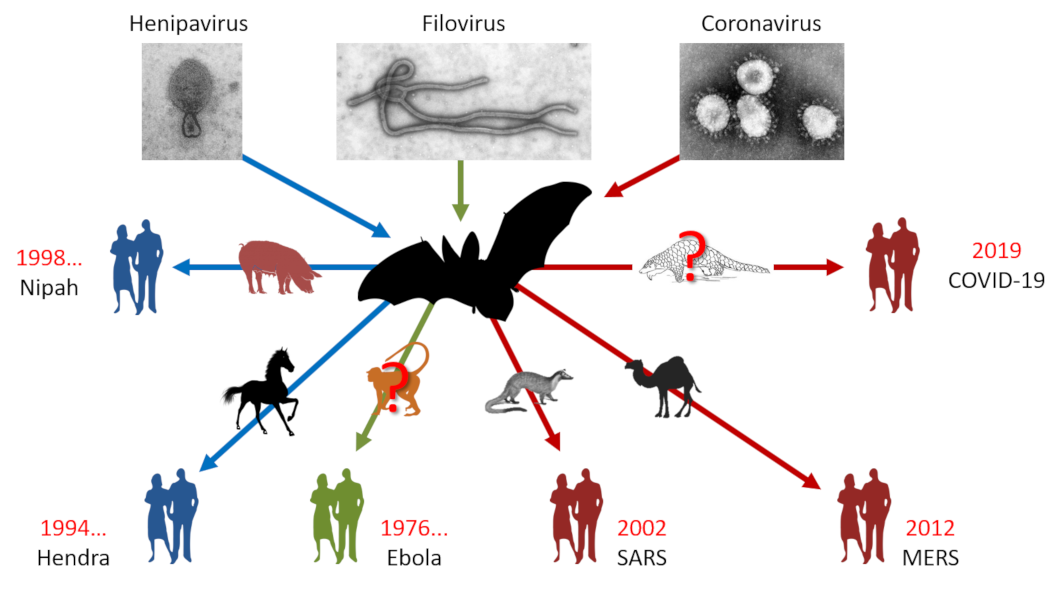
This is not the first time that bats have been in the spotlight. We have to go back to 1911 for the first mention of a virus isolated from bats. At the time, rabies was the subject of interest and it was discovered that it was transmitted by the common vampire bats of Brazil. After a long period of oblivion, it was rediscovered during the years 1990-2000 in the framework of health surveillance programmes that bats are the natural hosts of many emerging viruses such as henipaviruses (Hendra and Nipah), filoviruses (Ebola) and coronaviruses (SARS) [3] (Figure 1). Unexpectedly, they are found to be unaffected by most of these pathogens. It has even been demonstrated, particularly for the Hendra and Nipah viruses, that they can withstand viral loads that are normally lethal in other mammals [4].

2. Bats and viruses?

2.1. Bats account for a quarter of all mammalian species

Figure 2. The order Chiroptera (bats) accounts for nearly a quarter of mammalian species. [Source: Aranae, Public domain, via Wikimedia Commons]
Bats are the only flying mammals grouped in the order Chiroptera, whose name comes from the Greek kheir, “hand” and pteron, “wing” meaning “that flies with its hands”. This order includes a large number of species currently estimated at more than 1 428 or about a quarter of the 6 495 known mammalian species, placing it second only to the order of Rodents (Figure 2).

A recent revision of the order of Chiroptera based on molecular data has recast it into two new suborders that are more phylogenetically coherent (See: Focus Bats and Coronavirus Emergence) :

chauve souris - chauves souris - renards volants à lunettes
Figure 3. Spectacled flying foxes (Pteropus conspicillatus) in Cairns, Australia. They live mainly in the canopy of tropical rainforests. [Source: © François Moutou]
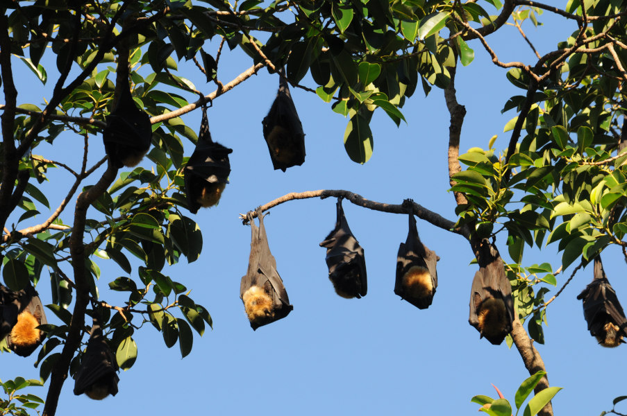
  • Yinpterochiroptera (Pteropodiformes), which includes tropical fruit bats or flying foxes (Figure 3);
  • The Yangochiroptera (Vespertilioniformes) which includes all microchiroptera except five families.

2.2. A very wide variety of ecological niches

chauve souris - chauves souris - chauves souris virus - chauve souris coronavirus - chauve souris covid19 - petit rhinolophe
Figure 4. Lesser horseshoe bat (Rhinolophus hipposideros) in flight. It prefers hedgerows made up of grazed meadows interspersed with wooded hedges. [Source: © Louis-Marie Préau]
Bats display an exceptional diversity of species that are widespread on all continents except the North and South Poles and occupy a very wide variety of ecological niches (Figures 3 & 4). As regards their diet, they are mainly frugivorous or insectivorous, but they may also be nectarivorous, pollinivorous, carnivorous (fish, amphibians, small mammals), omnivorous and haematophagous. This last diet concerns only three species of bats called vampires, located in tropical areas of the American continent. Although they rarely attack humans, these vampires are the object of unjustified fears among the public. Nevertheless, they can transmit the rabies virus by ingesting the blood of mammals. Thus, before going any further in the presentation, we can see that bats are a very favourable “breeding ground” for the multiplication, exchange and spread of viruses.

2.3 What are bat viromes?

Figure 5. Analysis of different bat viromes. [Source: Author’s figure]
The relatively recent rediscovery of bat/virus links will spark interest among researchers in this new way of approaching diseases from an ecological and health perspective. In addition to this interest, recent advances in investigative methods will be added to this craze, using metagenomic and high-throughput sequencing approaches that make it possible to analyse all the viral genomes (virome) contained in a biological sample in record time and without the need for culture steps. All of this will lead to an acceleration of study programmes whose objectives are to improve knowledge of bat viromes (Figure 5). Campaigns will thus be launched to collect samples as diverse as guano (faeces), urine, oral or anal swabs, and even organs in countries where bat sacrifice is authorized [5].

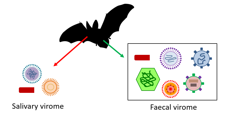
Thanks to these samples, a large number of viral sequences were identified, some of which were found to be similar to those of many viruses known to be pathogenic to humans [6]. Nevertheless, beware not to conclude, as has sometimes been done rather hastily, that bats are responsible for all the viral diseases that affect humans! Although these methods have brought significant advances in our knowledge of the links between viruses and bats, we must nevertheless remain cautious in interpreting the considerable mass of data obtained, given the weakness of the means of analysis.

However, it is surprising to note that all these advances have confirmed simple observations made in 1974, which already revealed the propensity of bats to host a wide variety of RNA and DNA viruses [7].

2.4. Are bats “virus strain”?

chauve souris - chauves souris - chauves souris virus - chauve souris coronavirus - chauve souris covid19 - virus chauve souris
Figure 6. Diversity of viruses found in bats. Coronaviruses are the most numerous, but studies are only just beginning. [Source: Diagram by Letko et al. [11] Copyright / link: doi: 10.1038/s41579-020-0394-z]
Although Rodents are the 1st order in number of species among Mammals, a comparative study [8] of the number of zoonotic viruses present in Rodents and Chiroptera has shown that their numbers are higher in Rodents, 68 versus 61, but that the ratio of the total number of viruses per species puts Chiroptera at the top (1.79 versus 1.48). More recently, a study by Peter Daszak and colleagues published in the journal Nature [9] showed that the total number of viruses infecting a mammalian species can be used to predict the proportion of viruses capable of causing zoonoses. One of the conclusions of this work is that bats proportionally harbour a higher number of zoonotic viruses than in all other orders of mammals.

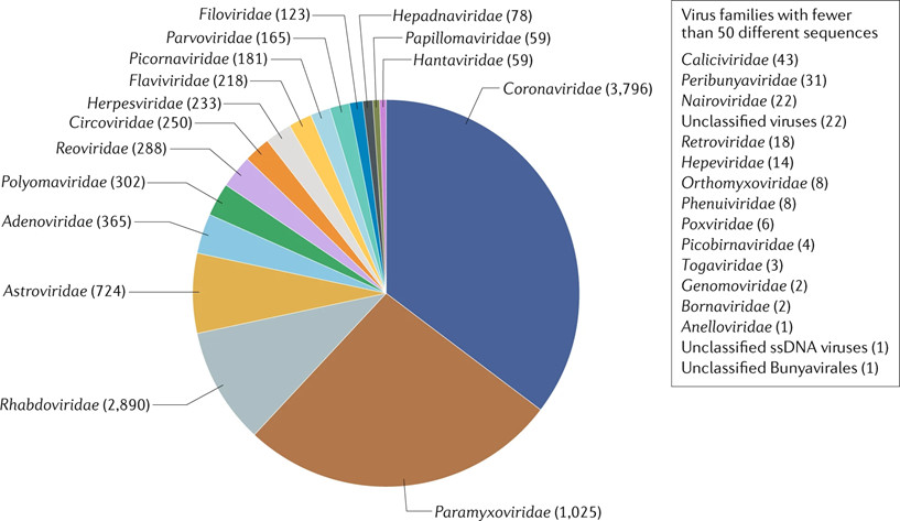
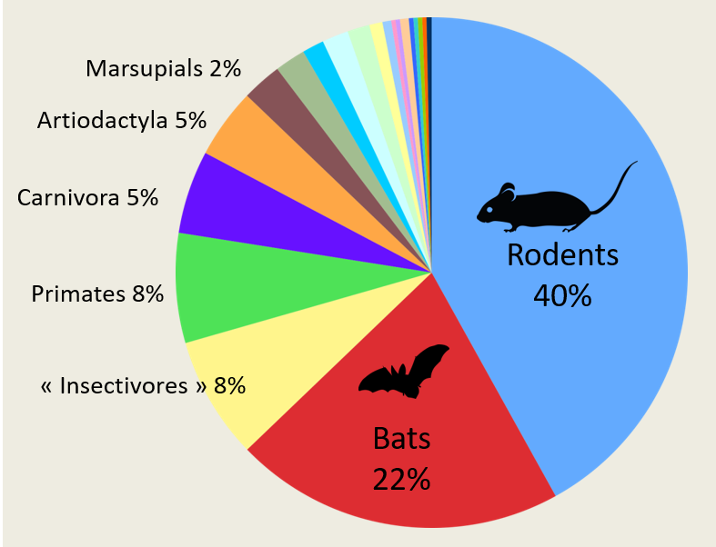
With regard to coronaviruses in particular, another large comparative study [10] was conducted in more than 19,000 animals belonging to bats, rodents and non-human primates. It showed that 98% of the coronaviruses found came from bats. A massive and diverse presence since, out of one hundred types of coronavirus identified, 91 came from Chiroptera. The proportion of individuals positive to a coronavirus was 8.6% in bats and 0.2% in other animals. Extrapolating these results to all species of bats, they would be carriers of no less than 3,200 coronaviruses throughout the world. This number was recently re-evaluated at 3,796 (Figure 6) [11]. Bats therefore seem to have special abilities.

Nevertheless, all this should be put into perspective because sampling bias exist in these studies. The order of Chiroptera, with its 1,428 species, occupies the second place among Mammals, and bats are inevitably at the forefront of the scene. Recently, researchers have shown that by going beyond mammals and including birds, the risk of viral zoonoses remains homogeneous regardless of whether the host species belong to the avian or mammalian orders, and thus the number of zoonoses per taxonomic order increases with species abundance [12]. Thus, for bats and rodents, the high number of zoonotic viruses is simply related to the specific richness of the two orders they represent.

In fact, explanations of the special role that bats play in viral emergences are more likely to be sought in terms of their particular ecoethology, their unusual physiological and immune characteristics, and also their ability to exchange RNA viruses more intensely than in other animal species. Finally, we must add the way in which humans, through their action on ecosystems, create favourable conditions for bringing all these elements into synergy.

3. Why are bats preferred hosts for pathogenic viruses?

3.1. Viruses and bats have been living together for a long time

fossile chauve souris
Figure 7. Age of bats. A, fossil of Onychonycteris finneyi, one of the oldest chiropteran fossils (Royal Ontario Museum) dating back 52.5 million years (Eocene). It was discovered in Wyoming, North America. This bat flew but was also able to climb trees. It hunted insects by using its sight and sense of smell as it was not yet equipped with the echolocation system. [Source: A, Matthew Dillon, CC BY 2.0, via Flickr]. B, probable representation of O. finneyi. [© N. Tamura; licence CC BY-SA]
The virus/bat cohabitation may have been established a very long time ago, since the oldest known fossil bats appeared with certainty in the Eocene more than 50 million years ago (Figure 7). Despite the Cretaceous-Tertiary biological crisis that occurred only 10 million years earlier, the diversity of bats in the Eocene is already remarkable. A recent publication [13] has confirmed the antiquity of the Chiroptera and shed new light on the origin of bats, which had until then been an enigma. By comparing the genomes of six bat species with those of other mammalian species, the researchers have shown that Chiroptera are indeed part of the laurasiatheran family, a super-order of placental mammals but that they share a common ancestor with the clade of Fereuungulata which groups together 5 orders (camels, pigs, cetaceans, horses, rhinoceroses, carnivores and pangolins) from which they have separated, pushing back the date of appearance of the Chiroptera to more than 65 million years.

3.2. A long period of co-evolution between viruses and bats

Viruses have thus had a very long time to adapt to their Chiropteran hosts and co-evolve intimately. A slow selection of the viruses’ capacities to infect their host took place, leading to increasingly close viral adaptation to certain molecules present on the surface of bat cells. Since these molecules act as receptors, they will play a crucial role in the penetration of the virus into the host cells. Moreover, being involved in a number of fundamental physiological processes, these receptor molecules have remained highly conserved in the animal world. This explains the ease with which the viruses play on species barriers and circulate rapidly among mammalian hosts.

Figure 8. After Spike protein binding of SARS-CoV-2 to the host cell receptor consisting of the angiotensin I converting enzyme 2 (ACE2) and activation by cleavage of Spike by a membrane protease, fusion of the viral complex takes place directly on the cell surface (A). In the absence of protease, the virus enters the cell by endocytosis (B). [Source: Author’s figure]
In the case of SARS-CoV-2, the receptor was rapidly identified as angiotensin I converting enzyme 2 (ACE2) (Figure 8). Involved in the regulation of blood pressure, is widely distributed in vertebrates, and is found on the cell surface of many tissues and organs (lungs, heart, arteries, kidney and digestive system). In infection, it acts as a lock and interacts with the key represented by the viral protein S (for Spike which means spicule). It is the spicules that give the virus its crowning glory, hence its name coronavirus.

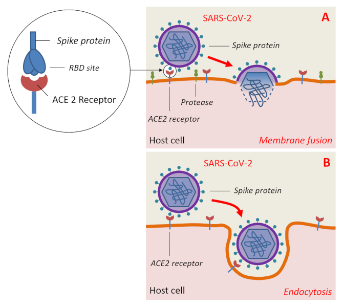
However, the key (S protein) must still have mutagenically acquired a specific amino acid sequence in its receptor binding domain (RBD) to the human ACE2 receptor as well as a specific site of cleavage by host enzymes so that SARS-CoV-2 may infect human cells [14].

These mutations were presumably acquired by the bat coronavirus during passage through intermediate hosts to result in SARS-CoV-2. In addition to humans, the latter is capable of infecting other mammals such as primates (Macaque monkeys), certain species of deer and cetaceans, domestic cats, tigers, golden hamsters, ferrets, mink and domestic dogs. Apart from Mammals, other classes of vertebrates do not seem to be affected [15].

3.3. Flying, essential to the links between bats and viruses

chauve souris - grand rhinolophe
Figure 9. Greater horseshoe bat (Rhinolophus ferrumequinum) in flight. [Source: © Louis-Marie Préau]
Among the adaptations that have made the success of the Chiroptera, the acquisition of flight during evolution is undoubtedly the most remarkable (Figure 9). It appears that flight played an essential role in the links between bats and viruses. In addition to the fact that it is an excellent means of movement for bats, it will contribute to the spread of viruses over large areas and in very varied ecosystems. For example, the geographical distribution of viruses of the genus Henipavirus has been found to overlap closely with that of bats of the genus Pteropus, with animal and human henipaviruses having been reported in Australia, Bangladesh, India, Malaysia and Singapore.

More unexpectedly, it was realized that flight, by greatly increasing the metabolism (15 to 16 times compared with the 2 times of most birds) and the bat’s body temperature (>38°C), induced a response similar to that of a feverish state. However, fever is a natural defence mechanism used by endothermic animals to inhibit the growth of pathogens and stimulate their immunity. Thus, by inducing a more or less permanent feverish state, flight would contribute to making bats more resistant to viral attacks [16].

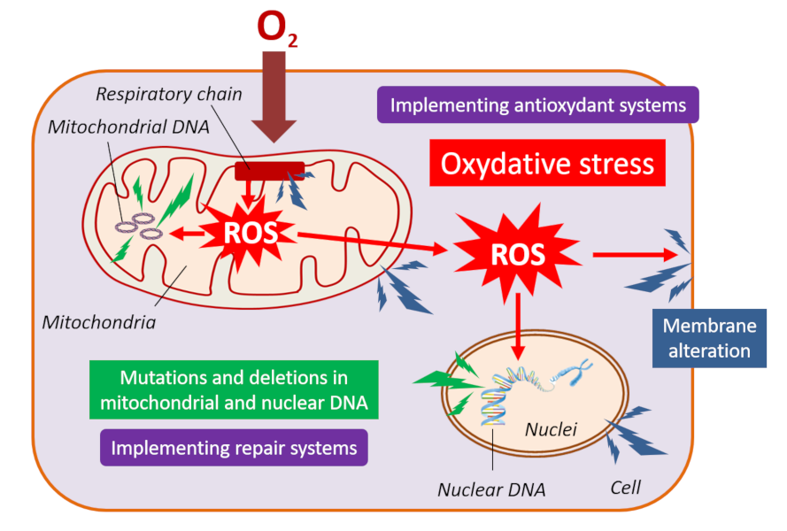
3.4. Breathing and oxidative stress

Figure 10. The production of reactive oxygen species (ROS) in the mitochondrial respiratory chain induces oxidative stress responsible for cell level alterations in membrane lipids, proteins and DNA. To protect itself, the cell has antioxidant and repair systems. [Source: Author’s figure]
During flight, bats consume four times more oxygen, which generates a large quantity of reactive oxygen species (ROS) at the level of the mitochondrial respiratory chain, causing intense oxidative stress (Figure 10). The latter usually causes significant damage to cellular constituents, including DNA, but bats seem to escape it. In fact, bats have selected particularly effective mitochondria and antioxidant systems to support the high energy demand required by flight and protect themselves from the effects of oxidative stress [17]. When the damage is done, bats can rely on a high-performance DNA repair system [18]. Since many pathogens generate oxidative stress in the initial stages of infection and mitochondria have an increasingly recognized role in the immune system, these adaptations, which were slowly selected by Chiroptera over the course of evolution, have had beneficial effects on their immunity and pathogen control [19].

3.5. Exceptional longevity conducive to virus persistence

Figure 11. Longevity quotient (ratio of observed to estimated longevity) for 779 mammalian species as a function of body weight (in grams). The black dashed line indicates a quotient = 1. Most bats have a high longevity as a function of their body mass (blue dots), as in the case of one rodent species, the naked mole rat (black star). The relationship between telomere length and age was estimated for bat species represented by silhouettes. [Source Foley et al. [20], CC BY-NC 4.0 / Link: https://advances.sciencemag.org/content/4/2/eaao0926]
At the same time, the viruses would have gradually become accustomed to their chiropteran host and would have had to lower their virulence level. In other words, everyone learned to live together harmoniously. Thus, it can be seen that many life traits in bats are conducive to the persistence of viruses.

We saw earlier that the bat manages to moderate oxidative stress, which generally leads to chronic inflammation and accelerated aging. Similarly, the progressive shortening of telomeres (chromosome ends), which normally causes senescence and the appearance of cancer, has not been observed in bats [20]. Bats thus show exceptional longevity (Figure 11) with few signs of senescence and a negligible rate of cancer. Out of 19 mammalian species that live proportionally longer than humans, taking their body size into account, 18 are bats, with a record held by a Brandt’s bat (Myotis brandtii), which, at a weight of 7 g, has exceeded the age of 41 years. This longevity is conducive to the persistence of viruses in individuals and to long-term viral exchanges between generations of juveniles and adults.

3.6. Lethargy and torpor

chauve souris hibernation - grand rhinolophe hibernation
Figure 12. Greater horseshoe bat in hibernation. A, colony of Greater horseshoe bats; B, Greater horseshoe bat (Rhinolophus ferrumequinum) in lethargy enveloped in its wings (we can see the nasal leaf which allows the concentration of ultrasounds emitted by the nostrils for echolocation deployed in active phase). [Source: © Marc Pihet]
Another factor favourable to the persistence of viruses concerns the states of lethargy or torpor into which Chiroptera can enter (Figures 12 & 13).

Lethargy is a strategy used by bats in temperate countries to survive the insect shortage that occurs during cold seasons while other animals such as birds choose to migrate to warmer climates.

petit rhinolophe lethargie - chauve souris lethargie
Figure 13. Lethargic Lesser horseshoe bat (Rhinolophus hipposideros) passively hanging from the wall by its claws. [Source: © Marc Pihet]
Thus during hibernation, which lasts several months, the bat falls into a state of profound lethargy that is accompanied by hypothermia and an extreme slowing of its metabolism in order to save energy. A weakening of the immune defences is associated; it is quite favourable to the maintenance of viruses. This explains the inter-seasonal transmission of viruses. As for tropical bats that do not hibernate, it has been observed that they periodically fall into states of daytime torpor, which are accompanied by a reduction in their metabolism as during hibernation [17].

3.7. Importance of living together

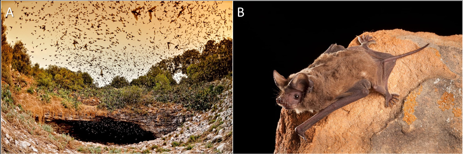
Viral persistence can also be expressed on an individual level, as well as on a collective level. Chiropterans have a strong propensity to group together in colonies of hundreds or even thousands of individuals of different ages and sometimes of different species. The largest known concentration of mammals is found in the Braken Cave in Texas, which concentrates more than 20 millions of Brazilian free-tailed bats (Tadarida brasiliensis mexicana) (Figure 14). It takes at least 3 hours for the entire colony to emerge from the cave!

bracken cave
Figure 14. A, Bracken Cave (Texas, USA) which hosts one of the largest bat colonies in the world with an estimated 20 millions of Brazilian free-tailed bats gathered from March to October. B, Brazilian free-tailed bat (Tadarida brasiliensis) [Source: A, photo Jonathan-Alonzo, photo courtesy of Bat Conservation International / B, Michael Durham Minden, photo courtesy of Bat Conservation International].
During these aggregation phenomena, it is obvious that viruses are transmitted very easily between bats, either by mutual licking of their hairs, which are frequently soiled by urine or faeces, or by aerosolisation of biological liquids (urine, saliva), which are good carriers for viral diffusion. To move around or hunt at night, some bats use echolocation (See Focus: Echolocation) which mobilizes the ear, nose and throat and generates aerosols from secretions of the nasal mucosa and saliva, the preferred sites for viruses. This faculty acquired during evolution consists of emitting ultrasounds to create sound “images” in an environment without light.

In these groupings, it has been well shown that the nature and size of colonies play an important role in viral maintenance and the appearance of new variants. It is thought that viral exchanges between adults and juveniles promote chronicity of infection, whereas between individuals of different species, viral diversity increases. Researchers are now wondering whether this habit of living in sympatry may be the determining factor that has led bats to tolerate viruses while developing unique immune capacities, aspects of which have only recently begun to be identified.

3.8. Unique immune abilities

Figure 15. Flying induces in bats unique immune capacities designed to control the viral attack and the resulting inflammatory reaction. [Source: Author’s Figure]
Normally, humans and most other mammals respond to a viral attack by activating their immune system at the time of infection. This is not the case in bats, which maintain this system permanently but at low noise levels so that it can quickly become more powerful in the event of infection (Figure 15). In fact, an initial inflammatory response makes it possible to send cytokines (interferons) that target the pathogenic agent as soon as it arrives, followed by a second response in which interleukins are used to temper the inflammation in order to prevent its deleterious effects on the host. In fact, it is flight, an intense and very pro-inflammatory activity, that is thought to have induced this unique ability to prevent the onset of exacerbated inflammation [18].The latter corresponds to the famous cytokine storm evoked during the COVID-19 pandemic, which provokes a disproportionate inflammatory reaction in some patients and which resuscitators try to control, like bats, because it threatens the patient’s vital prognosis [21].

In fact, bats seem capable of restricting their immune response to pathogens with which they share a long evolutionary history in order to limit the immunopathological consequences of an infection [22]. A kind of non-aggression pact seems to have been established between them and viruses.

Recent analysis of six bat genomes [13] has shown a wide variety of endogenous viruses, which tends to confirm this state of tolerance towards viruses recognised as elements of the bat self. It also revealed the concentration of the inflammatory response on a limited number of genes and the existence of new genes involved in tolerance to viral infections. This study aims to better understand how bats tolerate coronavirus infections.

4. Are bats responsible for viral outbreaks?

The recognition of the role of bats in viral epidemics presents the risk of bats being responsible for them and thus of considering their eradication as a solution to the risk of infection. This reaction, which is unfortunately intuitive, would prove to be totally inappropriate and even prejudicial to human health. It has already been tested in Uganda where, as part of campaigns to prevent Marburg virus infections, destruction of fruit bats has been carried out in some mines. This has resulted in re-invasion of these sites by susceptible bats and multiple reintroductions of the virus into new connected populations.

Reacting like this means forgetting that Chiropterans are key species in the functioning of ecosystems. In tropical environments, they play a significant role in the pollination of plants and the long-distance dissemination of seeds. Humans benefit by consuming them and using their guano as fertilizer. As for insectivorous bats, they play a major role in regulating insect populations and thus contribute to a reduction in the use of pesticides. Very sensitive to changes in their environment, bats are excellent indicators of the health of our environment.

Figure 16. Factors favouring zoonoses. [Source: Author’s figure]
The causes of epidemics are rather to be found in the disruption of natural ecosystems inflicted by human activities: intensification of agricultural practices leading to deforestation and habitat fragmentation, habitat degradation and rapid urbanization [23] (Figure 16). These activities encroach on areas that have long remained wild and bring bat populations closer to human habitats and domestic animal farms [24]. In addition, there is the unnatural grouping under deplorable conditions of live wild or domestic animals intended for sale on the markets, which are veritable cauldrons of emerging viruses. To avoid this, an easy and quick measure would be to apply strict controls on these live animal markets. In the longer term, human beings should be encouraged to make profound changes in their eating habits.

In a more comprehensive way, it will be necessary to act on the risk factors that expose humans to wildlife and bats. The COVID-19 pandemic is there to remind us of this. It made it clear that controlling these risks requires close collaboration between the fields of human health, animal health and ecosystem health. In other words: doctors, pharmacists, veterinarians and ecologists, as well as economists and lawyers, must work together to promote an ecology of health, an essential approach to prevent future epidemics and prevent them from turning into health crises (See Viral pandemics of the modern era).

5. Messages to remember

  • Bats are natural hosts for many emerging viruses, but are unaffected by most of them.
  • Bats are the preferred hosts of viruses because of their diversity of species, 2nd order in Mammals, diets, and ecological niches.
  • Research is being conducted on the virome (set of viral genomes) of bats, which shows that they are not directly responsible for human viral diseases.
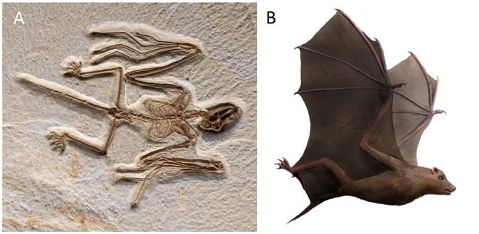
  • Their appearance dates back more than 65 million years, which has given them time to co-evolve closely with viruses.
  • The success of Chiropterans is due to their unique adaptations: flight and echolocation abilities, exceptional longevity, ability to enter states of lethargy or torpor, gregarious instinct and unique immune system that have contributed to their wide tolerance to infections
  • Chiropterans are key species in the functioning of ecosystems and excellent indicators of the health of our environment.
  • The causes of epidemics are rather to be found in the disruption of natural ecosystems due to uncontrolled human activities.
  • To prevent new epidemics, it is imperative to act on the risk factors that expose humans and wildlife.


Thanks to François Moutou for his proofreading of the article and his exotic photographs, and to Marc Pihet and Louis-Marie Préau (www.louismariepreau.com) for their photographic contributions illustrating this article.


Notes and References

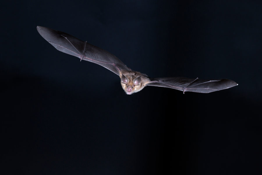
Cover image. Greater mouse-eared bat (Myotis myotis) in flight. [Source: © Louis-Marie Préau, www.louismariepreau.com]

[1] Zhou P, Yang XL, Wang XG, Hu B, Zhang L, Zhang W, Si HR, Zhu Y, Li B, Huang CL, Chen HD, Chen J, Luo Y, Guo H, Jiang RD, Liu MQ, Chen Y, Shen XR, Wang X, Zheng XS, Zhao K, Chen QJ, Deng F, Liu LL, Yan B, Zhan FX, Wang YY, Xiao GF & Shi ZL. (2020). A pneumonia outbreak associated with a new coronavirus of probable bat origin. Nature, 579 (7798), 270-273. <doi: 10.1038/s41586-020-2012-7>.

[2] Xiao K, Zhai J, Feng Y, Zhou N, Zhang X, Zou J-J, Li N, Guo Y, Li X, Shen X, Zhang Z, Shu F, Huang W, Li Y, Zhang Z, Chen R-A, Wu Y-J, Peng S-M, Huang M, Xie W-J, Cai Q-H, Hou F-H, Chen W, Xiao L & Shen Y. (2020). Isolation of SARS-CoV-2-related coronavirus from Malayan pangolins. Nature, 583 (7815), 286-289. <doi: 10.1038/s41586-020-2313-x>.

[3] Calisher CH, Childs JE, Field HE, Holmes KV & Schountz T. (2006). Bats: important reservoir hosts of emerging viruses. Clin. Microbiol. Rev., 19 (3), 531-545. <doi:10.1128/CMR.00017-06>.

[4] Middleton DJ, Morrissy CJ, van der Heide BM, Russell GM, Braun MA, Westbury HA, Halpin K & Daniels PW. (2007). Experimental Nipah virus infection in pteropid bats (Pteropus poliocephalus). J. Comp. Pathol., 136 (4), 266-272. <doi: 10.1016/j.jcpa.2007.03.002>.

[5] Hu D, Zhu C, Wang Y, Ai L, Yang L, Ye F, Ding C, Chen J, He B, Zhu J, Qian H, Xu W, Feng Y, Tan W & Wang C. (2017). Virome analysis for identification of novel mammalian viruses in bats from Southeast China. Sci. Rep. 7 (1), 10917. <doi: 10.1038/s41598-017-11384-w>.

[6] Drexler JF, Corman VM, Müller MA, Maganga GD, Vallo P, Binger T, Gloza-Rausch F, Rasche A, Yordanov S, Seebens A, Oppong S, Adu Sarkodie Y, Pongombo C, Lukashev AN, Schmidt-Chanasit J, Stöcker A, Carneiro AJ, Erbar S, Maisner A, Fronhoffs F, Buettner R, Kalko EK, Kruppa T, Franke CR, Kallies R, Yandoko ER, Herrler G, Reusken C, Hassanin A, Krüger DH, Matthee S, Ulrich RG, Leroy EM & Drosten C. (2012). Bats host major mammalian paramyxoviruses. Nat. Commun., 3, 796. <Law: 10.1038/ncomms1796>.

[7] Sulkin SE & Allen R. (1974). Virology. Vol. 8, Basel: Melnick JL, Karger.

[8] Luis AD, Hayman DT, O’Shea TJ, Cryan PM, Gilbert AT, Pulliam JR, Mills JN, Timonin ME, Willis CK, Cunningham AA, Fooks AR, Rupprecht CE, Wood JL & Webb CT. (2013). A comparison of bats and rodents as reservoirs of zoonotic viruses: are bats special? Proc. Biol. Sci. 280 (1756), 20122753. <Law: 10.1098/rspb.2012.2753>.

[9] Olival KJ, Hosseini PR, Zambrana-Torrelio C, Ross N, Bogich TL & Daszak P. (2017). Host and viral traits predict zoonotic spillover from mammals. Nature, 546 (7660), 646-650. <doi: 10.1038/nature22975>.

[10] Anthony SJ, Johnson CK, Greig DJ, Kramer S, Che X, Wells H, Hicks AL, Joly DO, Wolfe ND, Daszak P, Karesh W, Lipkin WI, Morse SS; PREDICT Consortium, Mazet JAK & Goldstein T. (2017), Global patterns in coronavirus diversity. Virus Evol. 3 (1), vex012. <Law: 10.1093/ve/vex012>.

[11] Letko M, Seifert SN, Olival KJ, Plowright RK & Munster VJ. (2020). Bat-borne virus diversity, spillover and emergence. Nat. Rev. Microbiol. 18 (8), 461-471. <doi: 10.1038/s41579-020-0394-z.>.

[12] Mollentze N & Streicker DG. (2020). Viral Zoonotic Risk Is Homogenous Among Taxonomic Orders of Mammalian and Avian Reservoir Hosts. Proc. Natl. Acad. Sci. USA, 117 (17), 9423-9430. <doi: 10.1073/pnas.1919176117>.

[13] Jebb D, Huang Z, Pippel M, Hughes GM, Lavrichenko K, Devanna P, Winkler S, Jermiin LS, Skirmuntt EC, Katzourakis A, Burkitt-Gray L, Ray DA, Sullivan KAM, Roscito JG, Kirilenko BG, Dávalos LM, Corthals AP, Power ML, Jones G, Ransome RD, Dechmann DKN, Locatelli AG, Puechmaille SJ, Fedrigo O, Jarvis ED, Hiller M , Vernes SC, Myers EW & Teeling EC. (2020). Six reference-quality genomes reveal evolution of bat adaptations. Nature, 583, 579-584. <doi: 10.1038/s41586-020-2486-3>.

[14] Zhang T, Wu Q & Zhang Z. (2020). Probable Pangolin Origin of SARS-CoV 2 Associated with the COVID-19 Outbreak. Curr. Biol. 30 (7), 1346-1351.e2. <doi: 10.1016/j.cub.2020.03.022>.

[15] Damas J, Hughes GM, Keough KC, Painter CA, Persky NS, Corbo M, Hiller M, Koepfli KP, Pfenning AR, Zhao H, Genereux DP, Swofford R, Pollard KS, Ryder OA, Nweeia MT, Lindblad-Toh K, Teeling EC, Karlsson EK & Lewin HA. (2020). Broad host range of SARS-CoV-2 predicted by comparative and structural analysis of ACE2 in vertebrates. Proc. Natl. Acad. Sci U.S.A. 2020 Aug 21:202010146. <doi: 10.1073/pnas.2010146117>.

[16] O’Shea TJ, Cryan PM, Cunningham AA, Fooks AR, Hayman DTS, Luis AD, Peel AJ, Plowright RK & Wood JLN. (2014). Bat flight and zoonotic viruses. Emerg. Infect. Dis. 20 (5): 741-745. <doi:10.3201/eid2005.130539>.

[17] Brook CE & Dobson AP. (2015). Bats as special reservoirs for emerging zoonotic pathogens. Trends Microbiol. 23 (3), 172-180. <doi: 10.1016/j.tim.2014.12.004>.

[18] Zhang G, Cowled C, Shi Z, Huang Z, Bishop-Lilly KA, Fang X, Wynne JW, Xiong Z, Baker ML, Zhao W, Tachedjian M, Zhu Y, Zhou P, Jiang X, Ng J, Yang L, Wu L Xiao J, Feng Y, Chen Y, Sun X, Zhang Y, Marsh GA, Crameri G, Broder CC, Frey KG, Wang L-F & Wang J. (2013). Comparative analysis of bat genomes provides insight into the evolution of flight and immunity. Science, 339 (6118), 456-460. <doi: 10 .1126/science.1230835>.

[19] Banerjee A, Baker ML, Kulcsar K & Misra V, Plowright R, Mossman K. (2020). Novel insights into immune systems of bats. Front. Immunol. 11, 26. <doi: 10 .3389/fimmu.2020.00026>.

[20] Foley NM, Hughes GM, Huang Z, Clarke M, Jebb D, Whelan CV, Petit EJ, Touzalin F, Farcy O, Jones G, Ransome RD, Kacprzyk J, O’Connell MJ, Kerth G, Rebelo H, Rodrigues L, Puechmaille SJ & Teeling EC. (2018). Growing old, yet staying young: The role of telomeres in bats’ exceptional longevity. Sci. Adv. 4 (2), eaao0926. <10.1126/sciadv.aao0926>.

[21] Kacprzyk J, Hughes GM, Palsson-McDermott EM, Quinn SR, Puechmaille SJ, O’Neill LAJ & Teeling EC. (2017). A potent anti-inflammatory response in bat macrophages may be linked to extended longevity and viral tolerance. Acta Chiropter, 19 (2), 219-228. <doi: 10.3161/15081109ACC2017.19.2.001>.

[22] Mandle JN, Schneider C, Schneider DS, Baker ML. (2018). Going to bat(s) for studies of disease tolerance. Front Immunol, 9 (2112). < doi: 10.3389/fimmu.2018.02112>.

[23] Gibb R, Redding DW, Chin KQ, Donnelly CA, Blackburn TM, Newbold T & Jones KE. (2020). Zoonotic host diversity increases in human-dominated ecosystems. Nature, 584 (7821), 398-402. <doi: 10.1038/s41586-020-2562-8>.

[24] Afelt A, Frutos R & Devaux C. (2018). Bats, Coronaviruses, and Deforestation: Toward the Emergence of Novel Infectious Diseases? Front. Microbiol. 9, 702. <doi: 10.3389/fmicb.2018.00702>.


环境百科全书由环境和能源百科全书协会出版 (www.a3e.fr),该协会与格勒诺布尔阿尔卑斯大学和格勒诺布尔INP有合同关系,并由法国科学院赞助。

引用这篇文章: LARCHER Gérald (2021年3月9日), Bats and viruses or how to live together in harmony, 环境百科全书,咨询于 2024年7月27日 [在线ISSN 2555-0950]网址: https://www.encyclopedie-environnement.org/en/life/bats-viruses-how-live-together-harmony/.

环境百科全书中的文章是根据知识共享BY-NC-SA许可条款提供的,该许可授权复制的条件是:引用来源,不作商业使用,共享相同的初始条件,并且在每次重复使用或分发时复制知识共享BY-NC-SA许可声明。