Symbiosis and evolution: at the origin of the eukaryotic cell

PDF
Encyclopedie environnement - eucaryote - eukaryotic cell

The cell of eukaryotic organisms (animals, plants, fungi) differs from that of prokaryotic organisms (Archaea and Bacteria) by the presence of several specialized organelles, such as: the nucleus (containing the genetic information of the cell), the mitochondria (site of cellular respiration), or the chloroplast (site of photosynthesis in plants). The existence and organization of mitochondrial and chloroplast DNA, as well as their biochemistry and some structural traits, have led to their being considered as ancient bacteria integrated into a host cell by an endosymbiosis process. One possible hypothesis would be that current eukaryotes would descend from an archaeal ancestor who acquired a proteobacteria, the present mitochondria. Once this step was established, some cells would have incorporated cyanobacteria that are the origin of the chloroplast. At the same time, they have acquired the ability to carry out photosynthesis, and thus an autotrophic metabolism, a particularity of plants. Throughout the process, gene transfer phenomena between symbionts, the taking over of the coding of some organelle proteins by the nucleus and the relocation of gene products into the organelles have closely integrated these prokaryotes within the host cell. The phenomenon of endosymbiosis is therefore very largely responsible for the biodiversity of eukaryotes that appeared during evolution. Thus, photosynthesis has developed in a wide variety of organisms: red and green algae, green plants through primary endosymbiosis, brown algae and many other organisms through secondary or tertiary endosymbiosis.


1. The eukaryotic cell is a chimera

Figure 1. Diagram of the structure of a eukaryotic animal cell. The animal cell is compartmentalized, it contains an endomembrane system (nuclear envelope, Golgi apparatus, endoplasmic reticulum, vacuoles…), mitochondria (limited by a double membrane), a cytoskeleton bathed in the cytoplasm. The nucleus and mitochondria contain DNA. Ribosomes (protein synthesis machinery) are present in two forms: 70S in mitochondria and 80S, generally in association with the reticulum.

EukaryotesSingle-cell or multicellular organisms whose cells have a nucleus and organelles (endoplasmic reticulum, Golgi apparatus, various plastids, mitochondria, etc.) delimited by membranes. Eukaryotes are, along with bacteria and archaea, one of the three groups of living organisms. correspond to multicellular organisms (animals, plants, fungi) and some unicellular organisms (protozoa, for example). The main characteristic of the eukaryotic cell (Figure 1) is the existence of a nucleus (in prokaryotes, the genome is only very rarely surrounded by a membrane) surrounded by a cytoplasm containing many organelles, such as mitochondriaOrganelles of the cytoplasm of eukaryotic cells (plants, algae, animals). As the site of cellular respiration, mitochondria convert the energy of organic molecules from digestion (glucose) into energy that can be directly used by the cell (ATP) during the “Krebs cycle”. This reaction requires the presence of oxygen and releases CO2, so it plays an essential role in the carbon cycle. Mitochondria originate from a prokaryotic organism (α-proteobacteria) integrated into eukaryotic protocells 2 billion years ago. (where respiration, present in all eukaryotic cells, takes place) and chloroplastsOrganelles of the cytoplasm of photosynthetic eukaryotic cells (plants, algae). As a site of photosynthesis, chloroplasts produce O2 oxygen and play an essential role in the carbon cycle: they use light energy to fix CO2 and synthesize organic matter. They are thus responsible for the autotrophy of plants. Chloroplasts are the result of the endosymbiosis of a photosynthetic prokaryote (cyanobacterium type) in a eukaryotic cell about 1.5 billion years ago. (site for photosynthesisBioenergetic process that allows plants, algae and some bacteria to synthesize organic matter from atmospheric CO2 by using sunlight. Solar energy is used to oxidize water and reduce carbon dioxide in order to synthesize organic substances (carbohydrates). The oxidation of water leads to the formation of O2 oxygen found in the atmosphere. Photosynthesis is the basis of autotrophy, it is the result of the integrated functioning of the chloroplast within the cell., in plants in the broad sense, terrestrial plants and algae). These organelles are frequently displaced or reorganized by the cytoskeleton that triggers intracellular mobility (Figure 1).

The eukaryotic nucleus is delimited by a double membrane called the nuclear envelope (Figure 1). It contains the nuclear genome characteristic of the eukaryotic cell, i.e. the genetic material of an individual encoded in its DNA (deoxyribonucleic acid). It is usually this genome that is referred to when the genome of a eukaryote is mentioned. However, the eukaryotic cell also contains non-nuclear genomes within the organelles:
– the mitochondrial genome, within the mitochondrial matrix (Figure 1);
– the chloroplastic genome, within the chloroplast stroma (e.g. plants or algae).

The DNA constituting these three genomes is not organized in the same way. In the nucleus, the genome is distributed over several linear DNA molecules, and organized into well-differentiated chromosomes. DNA contains all coding sequences (transcribed into messenger RNA, mRNA, and translated into proteins) and non-coding sequences (not transcribed, or transcribed into RNA, but not translated). The three-dimensional configuration of the nuclear genome has a functional importance: the winding (or “condensation”) of DNA on itself and around proteins, the histonesBasic proteins associating with DNA to form the basic structure of chromatin. Histones play an important role in DNA packaging and folding, allowing a large amount of genetic information to be packaged in the tiny nucleus of a cell. Mitochondrial or chloroplastic DNA do not have the same organization: it is generally circular, rarely linear (plant mitochondria), generally without intron, and is not associated with histone proteins.

ProkaryoticMicroorganisms (usually unicellular) with a simple cellular structure, no nucleus, and almost never internal compartmentalization (the only exception being thylakoids in cyanobacteria). Two of the three groups that make up living organisms are prokaryotes: Archaea and Bacteria.-type cells (Bacteria and ArchaeaSingle-celled prokaryotic microorganisms living in particular in extreme environments (anaerobic, high salinity, very hot…). Phylogenetic research by Carl Woese and George E. Fox (1977) differentiated between archaea and other prokaryotic organisms (bacteria). Currently, living organisms are considered to consist of three groups: Archaea, Bacteria and Eukaryota.), do not have a nucleus and their DNA is circular (or -in some rare cases- linear) and organized like that of chloroplasts or mitochondria. In this way, DNA replication, transcription and translation directly take place into the cytoplasm. It should be noted, however, that Archaea are only superficially similar to Bacteria in their cellular aspect: their metabolism differs greatly, and the mechanisms and proteins involved in the replication, transcription and translation processes have similar characteristics to those of eukaryotes. Finally, prokaryotes -in general- do not have internal compartments and, if present, they are less complex (cyanobacteria are an example of an exception). Above all, compartments, when they exist, are not mobile in the cell: the cytoskeleton, which is beginning to be discovered, does not move the cellular components within it.

Table 1. Comparison of eukaryotic and prokaryotic cells

Table 1 compares the properties of prokaryotic and eukaryotic cells (with their mitochondria and possibly their chloroplasts). It shows that mitochondria and chloroplasts have many characteristics in common with those of prokaryotic cells. Beyond the structure of DNA, eukaryotic cell organelles are formed from pre-existing organelles, dividing by fission to multiply, like a bacteria. Similarly, they have the same protein synthesis machinery (free 70S ribosomesA huge complex composed of RNAs and ribosomal proteins that allows the translation of mRNAs into proteins. Common to all cells (prokaryotes and eukaryotes), the ribosome varies according to the organisms: 80S ribosome in eukaryotes and 70S ribosome in prokaryotes and cellular organelles (mitochondria, chloroplast). in the matrix or stroma) while in the cytoplasm of the eukaryotic cell, this machinery consists of 80S ribosomes, sometimes fixed on the membranes of the endoplasmic reticulumMembrane network of the eukaryotic cell cytoplasm, essential for cellular metabolism (lipid and protein synthesis, calcium storage). Associated with ribosomes, it is the place of synthesis of proteins secreted outside the cell and, on the other hand, proteins and lipids constituting the membranes of cellular organelles (Golgi apparatus, lysosomes, mitochondria, nucleus, ribosomes, vesicles…).. Finally, bacteria also have the metabolism of mitochondria (i.e. respiration) and, in some peculiar cases, of chloroplasts (i.e. photosynthesis). On the other hand, the eukaryotic cell is distinguished by the existence of an active protein network, the cytoskeleton, a self-organized system capable of mobility, which positions and displaces the organelles in the cell. Such a protein network is static, or even absent, in prokaryotes, and poorly developed in mitochondria and chloroplasts.

Figure 2. Unrooted phylogenetic tree of the three domains of living organisms, produced using a gene from the small ribosomal subunit (bar: 0.1 substitution per site). The positions of the three genomes (nuclear, mitochondrial and chloroplastic) contained in maize (Zea mays) are indicated – Synechococcus is a cyanobacterium [Source: adapted from Ref. [1]]
The analysis of genome sequence by DNA sequencing techniques has provided information on the evolutionary history of living beings, including their relationship, also known as their phylogenyStudy of the links between related species. Allows to trace the main stages of the evolution of organisms from a common ancestor and to establish relationships of kinship between living beings. (see What is biodiversity? and Inheritance or convergence?…). Molecular phylogenetic analysis carried out on the nuclear genome of maize, as well as on its mitochondrial or chloroplastic genomes, makes it possible to determine the phylogenetic position of this plant within the tree of life (Figure 2). The analysis shows that three lines (two of which belong to Bacteria) are associated within what is considered, as they are structurally and functionally intertwined, as a single organism – showing the triple origin of the species.

All these properties show that the eukaryotic cell is a chimera containing both characteristic components of the eukaryotic cell (the nucleus) and organelles with typically prokaryotic properties (chloroplasts, mitochondria).

The distinction between Procaryotes and Eukaryotes was proposed in 1925 by Edouard Chatton [2] (who named these two cell types), although it was not recognized until the 1950s and 1960s. The chimeric nature of eukaryotic cells had been observed since the turn of the 19th to 20th centuries. If the botanist Andreas Schimper (born in France) had the idea in 1883 that photosynthetic organisms were the result of the combination of distinct organisms, it was the Russian biologist Constantin Mereschkowsky, who first provided solid arguments that some cells come from an intracellular union of two different types of cells (endosymbiosis). In his 1905 article [3], Mereschkowsky proposed three essential ideas: (a) chloroplasts resemble cyanobacteria that very early in the evolution established a symbiosis with a heterotrophic host, (b) the host that acquired the plastids was itself the product of an earlier symbiosis between a larger, heterotrophic, amoeboid-type host cell and a smaller, micrococcus-type endosymbionte that formed the nucleus, and (c) the autotrophy of plants is entirely inherited from cyanobacteria. Mereschkowsky had not considered the origin of the mitochondria. It is to the credit of the French microbiologist Paul Portier who wrote in a text in 1918 [4] that “all living beings, all animals (…), all plants (…) are constituted by the association, the interlocking of two different beings. Each living cell contains (…) formations that cytologists refer to as “mitochondria”. For me, these organelles would be nothing more than symbiotic bacteria, what I call symbionts. “These observations received no more attention from scientists than that, and the theory fell into disgrace, especially because plastids and mitochondria were not successfully cultured, which in the 19th century was considered as evidence of a bacterial nature [5]. It took new methods of studying the cell using electron microscopy, biochemistry and molecular biology for the theory of the endosymbiotic origin of organelles in the eukaryotic cell to be brought back to life around 1970 by the American microbiologist Lynn Margulis.

2. How did the eukaryotic cell evolved?

Figure 3. Hypotheses for the origin of eukaryotes. (A) In this so-called “Three Domains” scheme, the two Eukaryotic and Archaea lines have the same origin, each line being as old as the other. (B) The “two-domain” hypothesis comes from recent phylogenetic analyses.

Several hypotheses have been put forward to explain the appearance of the eukaryotic cell about 1.5 billion years ago, almost a billion years after the first prokaryotic organisms appeared on Earth. This question can be addressed in very different ways, depending on whether one considers palaeontological evidence, energy aspects, the origin of the characteristics of the eukaryotic cell or the relationships of the different prokaryotic and eukaryotic lines with respect to each other [6]. Figure 3 shows hypothesis for the origin of eukaryotes and other lines (Archaea and Bacteria).

Some models assume that eukaryotes emerged from a single ancestral lineage via successive mutations during the evolutionary process. Other models postulate that eukaryotes have emerged from a symbiotic association of prokaryotic cells whose fusion would have resulted in the transition from prokaryotic to eukaryotic. These various hypotheses can be partly tested by experience, in particular through the analysis of the genomes of current organisms (prokaryotes or eukaryotes) [7,8].

integration between the Archaean host cell and a α-proteobacteria
Figure 4. Schematic representation of the integration between the Archaea host cell and an α-proteobacteria to give a eukaryotic cell containing a mitochondria [see ref. 5]. (a-h) Illustrations of various steps describing the transition from an H2-dependent Archaea host cell (red) to an optional anaerobic protein bacteria (blue) to a eukaryotic cell. At first, the two organisms live nearby, the Archaea needs the hydrogen produced by the Bacteria, which does not support well the accumulation of hydrogen from its metabolism. The integration of the bacteria will follow and this transition is accompanied on one hand by gene transfer between the two organisms (c) and on the other hand by the establishment of the nucleus (e-h). [Source: reproduced with the permission of the authors (see ref. [5]) © 2015 (CC BY 4.0)]
The nature of the original host cell is a very controversial issue. The idea that it is an Archaea constitutes one of the possible scenarios for the appearance of the eukaryotic cell: according to this scenario, an Archaea host cell and a α-proteobacteriaA large group of bacteria called Gram-negative bacteria, because they have a cell wall rich in lipopolysaccharides and low in peptidoglycans. The mitochondria of current eukaryotic cells are thought to derive from one of these types of bacteria: alpha-proteobacteria. have established a stable symbiotic relationship (Figure 4, [5]). Among the many possibilities for establishing such a relationship [6], the existence of trophic relationships between two anaerobic prokaryotes living together, and drawing their food from this association (syntrophy), suggests an origin for mitochondrial symbiosis. In this hypothesis [5], the host would be an Archaea needing hydrogen in its environment to live (so-called H2-dependent Archaea which produces methane as metabolic waste) and the symbiote an optional anaerobic organism (α-proteobacteria) which could either breathe in the presence of O2 or perform H2-producing fermentations under anaerobic conditions. The latter metabolism produces energy only in low concentrations of H2 and benefits from the presence of H2-dependent archaea. Figure 4 shows how such a situation could have evolved into a eukaryotic cell [5, 9]. The strength of this hypothesis is that the partners need each other mutually, and that this scenario involves archaea and bacteria present in the current biosphere.

In this context, Bacteria and Archaea tend to interact closely in the same way as many current symbiotic associations. This can lead, in principle, to the situation described in Figure 4, in which the bacterial symbiote will be retained by the archeal host and eventually reside inside. In this case, the host does not feed on the symbiote, so integration is not due to a phenomenon of phagocytosisThe process by which a cell encompasses and then digests a foreign substance or organism (e g. bacteria) – although there is no doubt that phagocytosis has increased the frequency of integration of endosymbiotes in the evolution of eukaryotic cells [5].

The question of the origin of the eukaryotic cell is also linked to that of the nucleus, the emblematic structure of this cell. The establishment of a new membrane system, the nuclear membrane, in the host after mitochondria acquisition could be due to the aggregation of membrane vesicles composed of bacterial lipids. This separation between nucleus and cytoplasm could have responded to the need to separate, following gene transfer between host and symbiote, the splicing of RNA from DNA translation. It is then the selective pressure that would have led to the fixation of the compartmentalization between the newly formed nucleus and the cytoplasm [5] (Figure 4).

Thus, all eukaryotes currently known would descend from an archaean ancestor who acquired a proteobacterium during the Precambrian period, which became the mitochondria. This step is crucial: the integration of the mitochondria is therefore inseparable from the appearance of the eukaryotic cell as we know it today. The strong energy constraints exerted on the organization of prokaryotic cells were a major factor of innovation at the origin of the evolution of this cell. Only cells that had mitochondria had sufficient energy resources to reach the complexity of the eukaryotic cell, which is why there are no real intermediaries in the transition from prokaryotes to eukaryotes. It is often considered that it is only after this stage has been established that some of these cells have acquired the characteristics of the eukaryotic cell (nucleus, compartmentalization) and, eventually, integrated cyanobacteria. At the same time, they have acquired the ability to carry out photosynthesis, which is the origin of chloroplast, thus giving them an autotrophic metabolism.

Recently, some work has nuanced the age of mitochondrial symbiosis [10]. They are based on the age of acquisition of bacterial genes present in the eukaryotic nucleus (i.e. the date of their divergence from homologous genes currently found in free bacteria). They revealed that many genes, some of which contribute to the complexity of the eukaryotic cell, were most likely acquired before mitochondria. This does not imply that the cell into which the mitochondria entered was as complex as it is now, but it may have already been capable of phagocytosis, for example. This feature (specific to eukaryotes because it depends on the mobility of the cytoskeleton) could have helped in the placement of the mitochondria. The development of eukaryotic complexity therefore remains speculative, but may have begun before mitochondria, even if it undoubtedly benefited from it afterwards.

3. The endosymbiotic origin of the chloroplast

During phagocytosis in white blood cells or many protozoa (Figure 5), ingested cells are often directly digested (as in the case of prey), but sometimes they are permanently lodged in the cells (endosymbiotes). In the endosymbiosis process, organelle therefore results from internalization by phagocytosis without digestion of a prokaryote within a eukaryote (Figure 5). This is the case for chloroplasts in terrestrial plants, but also for red and green algae that are close to them [11,12].

Figure 5. Phagocytosis and primary endosymbiosis. During phagocytosis, ingested prey is often directly digested, but sometimes permanently lodged in the cells during primary endosymbiosis, the plasma membrane of the cell invades around the prokaryote and isolates it within an endocytosis vesicle. Then, when the prokaryote is integrated into the eukaryotic cell, the membrane of this vesicle disappears as well as the layer of peptidoglycans located between the two membranes of the cyanobacterium [see ref. 9 & 10].
During phagocytosis processes, the plasma membrane of the cell invades around the prey and isolates them into endocytosis vesicles where they are then digested as these vesicles fuse with others, the lysosomes, which contain enzymes. By analogy, it was generally considered that the outer membrane of the organelles came from this endocytosis membrane. Things are likely to be more complex (Figure 5). Indeed, the prokaryotes at the origin of chloroplasts or mitochondria are Gram- bacteria, characterized by the existence of a double membrane on the periphery of the bacteria. The outer membrane of chloroplasts, and in particular its outer surface immersed in the cell’s cytosol, contains characteristic glycolipids found in cyanobacteria [9, 10, 13]. It is therefore possible that the endocytosis membrane may have disappeared during the integration of the prokaryote into the eukaryotic cell. This is currently observed in Elysia chlorotica (see Focus), a marine mollusc that grazes algae, digests part of their cells but not the chloroplasts which is integrated into the cytoplasm of some of its cells. These chloroplasts remain functional throughout the life of the mollusc, which benefits from photosynthesis.

Primary and secondary endosymbioses
During the evolution, several endosymbiosis events repeated themselves and led to the formation of particular organisms. In primary endosymbiosis, the eukaryotic cell integrates a living prokaryotic. Thus, the chloroplasts of green line plants (red and green algae, to which terrestrial plants are attached) are derived from primary endosymbioses involving cyanobacteria. In some eukaryotes, mitochondria have evolved as a result of adaptation to anaerobic environments, but have never disappeared: they have produced particular mitochondria (hydrogenosomesOrganelles producing hydrogen, derived from a mitochondria. It is found in some anaerobic ciliates, Trichomonas, and fungi.) carrying out H2-producing fermentation (for example in some Ciliates) [14], but also small organelles, only involved in biosynthesis for the host cell, the mitosomesOrganelles present in some single-celled eukaryotic organisms, probably lacking DNA but with biosynthesis functions. [15].

Figure 6. Secondary chloroplastic endosymbiosis model in the cryptophyte Guillardia theta [see ref. 18]. Here, the nucleus of the internalised red algae (primary host) persists within the secondary host in the form of a vestigial nucleus (or nucleomorph), but with a very small genome (551 kb instead of the 350 Mb of the nucleus).
Secondary endosymbioses are a reiteration of the process, when a eukaryote already containing an endosymbiont realizes a secondary endosymbiosis within another eukaryote (Figure 6). This is the origin of plastids with more than two membranes present in some groups: internalisation of a green algae in Euglenes; independent internalisation of a red algae in brown algae, etc. Tertiary endosymbiosis, less frequent, have also been described. These various symbioses constitute as many founding endosymbioses of evolutionary lines [16,17].

4. Integration of the prokaryote into the eukaryotic cell

All these lines have a common characteristic: a strong genetic regression of endosymbiotes. Compared to free proteobacteria such as Escherichia coli, mitochondria have lost 99% of their genes. In the extreme, hydrogenosomes and mitosomes simply no longer have a genome! Green line plastids show a 95% genetic regression compared to single-celled free cyanobacteria: the number of genes has increased from several thousand in cyanobacteria to about 100 to 200 in chloroplasts… or even none in the regressed plastids of the parasitic plant Rafflesia.

The cause of this regression is obviously the loss of genes necessary for free living, or even for certain metabolic functions. For example, as with all Gram-bacteria detected by a staining technique called Gram staining: they then appear pink under the microscope. The staining technique is based on the membrane and wall characteristics of the bacteria. However, this is not a phylogenetic classification factor: indeed, the’Gram +‘ and’Gram ‘ groups are both non-monophyletic., a layer of peptidoglycancomposed of the wall of Gram positive and Gram negative bacteria. Consists of a carbohydrate part (= polysaccharide) and a peptide part. It maintains the shape of the cells and provides mechanical protection against osmotic pressure. is located between the two membranes of cyanobacteria, essential for maintaining the structure of bacteria in the natural environment, of low osmolarityNumber of moles of “osmotically active” particles in solution in 1 litre of solution. Concept related to the osmotic pressure exerted by the particles in solution, and responsible for osmosis. Sucrose is a small osmotically active molecule while starch is a huge osmotically inactive glucose polymer. The accumulation of sucrose in a compartment leads to an increase in osmotic pressure in that compartment, which is not the case with starch.. Once integrated into the host cell, the prokaryote will find itself in a medium, the cytoplasm, whose osmolarity is very close to that of its inner medium. The peptidoglycan layer then becomes useless, and the genes responsible for the placement of the peptidoglycan layer are then lost in chloroplasts (except in glaucophytesUnicellular plankton eukaryotes living in lakes, ponds or wetlands in temperate regions. They have flagella (2 of unequal length). They have chloroplasts (called cyanelles) that are blue-green in colour, due to the presence of phycocyanins and allophycocyanins in phycobilisomes. This is a group of reduced diversity.).

Although the organelle genome is regressing, the organelle protein repertoire (the proteome), when known, remains similar to that of the free bacteria proteome: proteins operating with new functions have therefore compensated for the losses. Their coding has in fact been supported by the host’s nuclear genome: genes located in the nucleus are translated into proteins in the cytosol which are addressed to the organelle through a transit peptidePeptide sequence located at the NH2-terminal end of the newly synthesized proteins in the cytoplasm and which allows them to be addressed to the specific organelle (mitochondria, etc.) where they function. We also speak of addressing peptide.. This phenomenon of relocation of the gene product into the organelle is an absolutely essential phenomenon for the integration of the prokaryote into the host cell. The addressing machinery responsible for these transfers is a converging innovation in plastids and mitochondria. It is also an example of the new functions linked to intracellular life. These machines, which allow the import of proteins synthesized in the cytosol through the two limiting membranes of mitochondria and chloroplasts, contain a large number of proteins whose evolutionary origin is complex: proteins of both prokaryotic and eukaryotic origin, encoded in the organelle and nucleus, are found there. Together, they allow the recognition of the protein being addressed, its unfolding followed by import (the protein must be maintained in an unfolded state to cross the membranes), then the cleavage of the addressing peptide before its precise location in its functional compartment [19].

What is the origin of the genes that encode in the nucleus for functions in organelles? There are actually two (Figure 7) [16]. Sometimes, original nuclear genes have replaced organelle genes: the corresponding gene product has acquired the ability to be addressed in the organelle. This activation may have in the past led to a redundancy situation whenever a gene already encoded the same function in the organelle. From this redundancy, the organelle gene could be lost without damage (Figure 7a) [16].

Figure 7. Evolutionary mechanisms leading to the replacement of organelle genes by genes located in the nucleus. Substitution (A) involves genes of “true” nuclear origin while transfer (B) involves a nuclear relocation of genes from organelles. [Source: According to Selosse et al (2001) Reference [16]]
Other cases involve gene transfers from organelle to the nucleus, which takes place in two major steps (Figure 7b). First, a DNA fragment encoding the organelle protein is relocated and then integrated into the nuclear genome. The transferred sequence will only code if, through mutations, it adapts to the nuclear genetic code, and if it acquires regulatory sequences for transcription. It must also acquire the pre-sequence corresponding to the transit peptide, which will ensure proper addressing of the mature protein to the organelle and therefore its correct location. As above, this leads to genetic redundancy: one or the other of the copies can be lost without damage. The loss of functionality and/or the disappearance of the organelle copy then seals the transfer (Figure 7) [16].

The passage of DNA fragments from organelles to the nucleus is not uncommon: large blocks of organelle DNA are inserted into the genome of some plants. These can be activated: nearly 10% of Arabidopsis thaliana‘s nuclear genes are thus derived from transfers from plastids, often followed by duplications [20]. It is not known how the DNA of the endosymbiote could have been integrated into the host genome, but it is assumed that this occurs during degradations of damaged or aged organelles accidentally releasing pieces of DNA into the host cytoplasm which are then randomly integrated into the host’s nuclear DNA.

The cytoplasmic genomes of organelles are at the crossroads of various selective forces, some of which favour their regression (such as the need for co-expression of certain genes), others favour the persistence of certain genes in the organelle genome. This could be the case for selection for a small genomic size that accelerates the multiplication of organelles and allows better transmission to daughter cells: it selects in particular the transfer of genes to the nucleus. The latter thus accumulates genetic potentialities from different lines coexisting with him in the cell [16]. Thus, while endosymbiosis reduces the genomes of endosymbiotes, it nourishes the genome of the host nucleus, contributing to its genetic diversification, and pushing for closer espouses the endosymbiotic association. Endosymbiosis therefore mixes the evolutionary lines present, by nesting but also by genetic chimerization in the nucleus of the host cell.

Finally, vertical transmission of the endosymbiote through generations is essential for the endosymbiosis to persist. Plastids must divide before the division of the host cell and must be half distributed in the two daughter cells. If their division is too rapid, they could take advantage of the host cell. On the contrary, a low division rate could lead to their disappearance. In this context, the establishment of coordination of cell division and symbiote division has been an essential element in the success of endosymbiosis. While most of the proteins involved in chloroplast division come from the cell division machinery present in cyanobacteria, some proteins are apparently of eukaryotic origin, and all are encoded in the nucleus: this is a way for the host to exercise control over chloroplast division.

5. Is symbiosis driving evolution?

Encyclopédie environnement - eucaryote - endosymbioses
Figure 8. Endosymbiosis (primary, secondary and tertiary) in the history of plastid evolution. They are responsible for organisms as diverse as red and green algae, terrestrial plants such as apicomplexes (parasites responsible for malaria and toxoplasmosis) or dinoflagellates (components of marine plankton that are particularly important in primary ocean production). [Source: reproduced from Keeling et al [12]. Copyright 2016 by American Journal of Botany, Inc]
In conclusion, extremely diverse symbioses that led to the formation of the eukaryotic cell [1,9], are at the origin of the development of eukaryotic biodiversity during evolution. Endosymbioses found new evolutionary lines. An example of the extreme diversity of organisms derived from endosymbiosis that causes chloroplasts is shown in Figure 8. However, things are not fixed: evolution continues to repeat itself! Today, some unicellular algae, cryptophytesSingle-celled organisms, mostly photosynthetic. Their chloroplasts are limited by four membranes, indicating an endosymbiosis of a photosynthetic eukaryote. Cryptophytes occur in many environments, particularly aquatic ones (oceanic environments, fresh waters, wetland pore waters). Some species have become intestinal parasites of metazoans. Some are Dinophyte endosymbiotes and heterocontes (Figure 8), whose four-membrane plastid derives from a secondary endosymbiosis, live in symbiosis in the cytoplasm of dinoflagellates that have lost their own plastids: there are three successive endosymbioses there!

More than a biological curiosity, symbiosis is certainly one of the most powerful motors of the evolution of the living world. It very quickly creates chimeric organisms that can generate new lines. It brings partners closer together and promotes massive gene transfers that also create chimeric genomes: the nuclear genome thus contains eukaryotic genes, but also genes of bacterial origin, derived from mitochondria, or even plastids, with which it borders. Such events may explain the major evolutionary leaps whose evolution seems to be punctuated, which have given rise to the great lines of life and shaped current biological diversity.

Thus, renewing the Darwinian vision of evolution by descent with modification, where one species is likely to give two, the endosymbiosis mechanisms remind us that sometimes two species, previously free and recognizable, merge into one. Man himself can be considered as an extremely integrated symbiotic community, formed by the eukaryotic cytoplasm and mitochondria, but also by the archaea and bacteria that populate, for instance, his gut microbiota…


References and notes

[1] Lang T. et al (2000) Autophagy and the cvt pathway both depend on AUT9. J Bacteriol 182, 2125-2133.

[2] Chatton E. (1938) Titres et travaux scientifiques (1906-1937). Sette, Sottano, Italy. L’histoire des conditions dans lesquelles Chatton a établi le concept de procaryote et eucaryote est décrite par Sapp J. (2005) The Prokaryote-Eukaryote Dichotomy: Meanings and Mythology, Microbiol Mol Biol Rev. 69, 292–305.

[3] Mereschkowsky C. 1905 Uber Natur und Ursprung der Chromatophoren im Pflanzenreiche. Centralbl. 25, 593-604; translated by Martin W, Kowallik K. (1999) Annotated English translation of Mereschkowsky’s 1905 paper ‘Uber Natur und Ursprung der Chromatophoren im Pflanzenreiche‘. Eur. J. Phycol. 34, 287–295.

[4] Portier P. (1918) Les Symbiotes. Masson (ed.), Paris. (in french)

[5] Martin W.F., Garg S. & Zimorski V. (2015) Endosymbiotic theories for eukaryote origin. Phil. Trans. R. Soc. B370, 20140330.

[6] Selosse M.A. (2012). Gloire et disgrâce de la théorie endosymbiotique. La Recherche 468: 92-94. (in french)

[7] Archibald J.M. (2014) One plus one equals one: symbiosis and the evolution of complex life. Oxford, UK: Oxford University Press.

[8] McFadden G.I. (2014) Origin and Evolution of Plastids and Photosynthesis in Eukaryotes, Cold Spring Harb.Perspect. Biol. 6, a016105

[9] Martin W. & Müller M. (1998) The hydrogen hypothesis for the first eukaryote. Nature 392, 37-41.

[10] Ettema T.J.G. (2016) Mitochondria in the second act. Nature 531, 39-40doi:10.1038/nature16876

[11] Archibald J.M. & Keeling P.J. (2002) Recycled plastids: a “green movement” in eukaryotic evolution. Trends Genetics 18, 577-584.

[12] Keeling P.J. (2004) Diversity and evolutionary history of plastids and their hosts. Am. J. Bot. 91, 1481-1493.

[13] Douce R., Block M.A., Dorne A.J., Joyard J. (1984) The plastid envelope membranes: their structure, composition, and role in chloroplast biogenesis. Subcell. Biochem. 10, 1-84, Springer US (Ed.)

[14] Selosse M.A. & Loiseaux-de Goër S. (1997) La Saga de l’endosymbiose, La Recherche 296, 36 (in french)

[15] Embley T.M. & Martin W. (2006) Eukaryotic evolution, changes and challenges. Nature 440, 623-630

[16] Lefèvre T., Renaud F., Selosse M.-A. & Thomas F. (2010). Évolution des interactions entre espèces, in F. Thomas, T. Lefèvre & M. Raymond (ed.), Biologie évolutive, p. 530-613. De Boeck, Paris. (in french)

[17] Keeling P.J. (2010) The endosymbiotic origin, diversification and fate of plastids. Phil. Trans. R. Soc. B 365, 729-748

[18] Douglas S. et al (2001) The highly reduced genome of an enslaved algal nucleus. Nature 410, 1091-1096.

[19] Selosse M.A., Albert B. & Godelle B. (2001) Small is successful: selection for reducing organelle’s genome size favours gene transfer to the nucleus. Trends Ecol Evol 16, 135-141.

[20] Jarvis P. (2004) Organellar Proteomics: Chloroplasts in the Spotlight. Current Biology 14, R317-9. http://www.cell.com/current-biology/references/S0960-9822%2804%2900231-3

Registering

Registering

Registering

Registering

Registering

Registering

Registering


The Encyclopedia of the Environment by the Association des Encyclopédies de l'Environnement et de l'Énergie (www.a3e.fr), contractually linked to the University of Grenoble Alpes and Grenoble INP, and sponsored by the French Academy of Sciences.

To cite this article: SELOSSE Marc-André, JOYARD Jacques (April 1, 2019), Symbiosis and evolution: at the origin of the eukaryotic cell, Encyclopedia of the Environment, Accessed July 27, 2024 [online ISSN 2555-0950] url : https://www.encyclopedie-environnement.org/en/life/symbiosis-and-evolution-origin-eukaryotic-cell/.

The articles in the Encyclopedia of the Environment are made available under the terms of the Creative Commons BY-NC-SA license, which authorizes reproduction subject to: citing the source, not making commercial use of them, sharing identical initial conditions, reproducing at each reuse or distribution the mention of this Creative Commons BY-NC-SA license.

共生与进化:真核细胞的起源

PDF
Encyclopedie environnement - eucaryote - eukaryotic cell

  真核生物(动物、植物、真菌)的细胞与原核生物(古菌和细菌)的细胞区别在于是否存在特异性的细胞器,如:细胞核(遗传信息储存的场所)、线粒体(细胞呼吸的场所)或叶绿体(植物光合作用的场所)。线粒体和叶绿体中存在DNA,具有独特的生物化学和结构特征,因此人们认为它们是古菌通过内共生作用逐渐融入宿主细胞中形成的。有一种可能成立的假设,即如今的真核生物起源于一个意外俘获了变形菌的古老祖先,而这个变形菌正是如今的线粒体。如果这一假设成立,还可能存在一些细胞意外融合了蓝细菌(蓝藻),进而形成叶绿体。同时,它们得到了进行光合作用的能力,实现了自养代谢——一项植物的特性。在整个过程中,共生体间的基因转移、细胞核对某些细胞器蛋白质的编码以及基因产物转移到细胞中,使得这些原核生物与宿主细胞紧密结合。因此,内共生现象是进化过程中真核生物多样性出现的主要原因。由此,光合作用得以在多种生物中进行:红藻和绿藻、通过初级内共生形成的绿色植物、褐藻和许多通过二次甚至三次内共生形成的其他生物。

1.真核细胞是嵌合体

环境百科全书-生命-真核动物细胞结构示意图
图1. 真核动物细胞结构示意图。
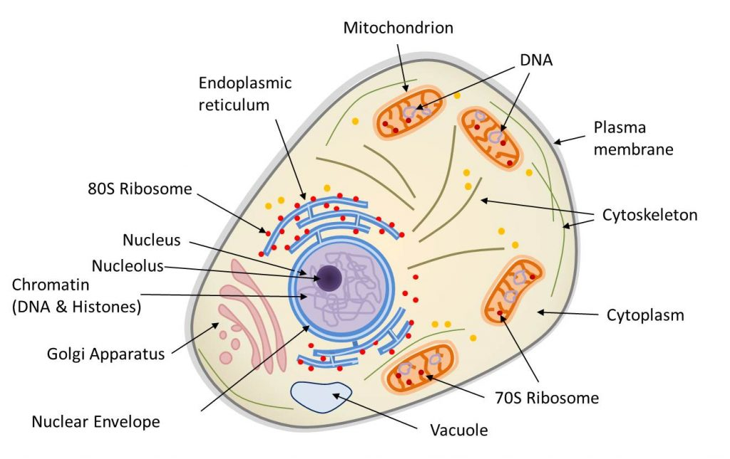
动物细胞是区室化的,由内膜系统(核膜、高尔基体、内质网、液泡等)、线粒体(受双层膜限制)和胞质内的细胞骨架组成。细胞核和线粒体都含有DNA。核糖体(蛋白质合成机器)以两种形式存在:70s核糖体(存在于线粒体中)和80s核糖体(通常与网状结构相关联)。(图1 Mitochondria 线粒体;Endoplasmic reticulum 内质网;80S Ribosome 80S核糖体;Nucleus 细胞核;Nucleolus 核仁;Chromatin 染色质;Histones 组蛋白;Golgi Apparatus 高尔基体;Nuclear Envelope 核膜;Vacuole 液泡;70S Ribosome 70S核糖体;Cytoplasm 细胞质;Cytoskeleton 细胞骨架;Plasma membrane 质膜)

  真核生物(单细胞或多细胞生物,其细胞拥有由膜界定的细胞核和细胞器(内质网、高尔基体、各种质体、线粒体等)。真核生物与细菌和古菌构成了生物的三种类型)包括多细胞生物(动物、植物、真菌)和单细胞生物(如原生动物)。真核细胞(图1)的主要特征是存在细胞核(在原核生物中,基因组很少被膜包围)、细胞质中含有许多细胞器,如线粒体【核细胞(植物、藻类、动物)胞质器:作为细胞呼吸的场所,线粒体通过“克雷布斯循环”将消化产生的有机分子(葡萄糖)的能量转化为细胞直接使用的能量(ATP)。这一反应需要氧气的存在并释放CO2,因此在碳循环中发挥着重要作用。线粒体起源于20亿年前融入真核原细胞的原核生物(α-变形菌)。】(存在于所有真核细胞中,负责呼吸作用)和叶绿体【进行光合作用的真核细胞(植物、藻类)的胞质器:叶绿体作为光合作用场所,产生氧气(O2)并在碳循环中发挥着至关重要的作用:它们利用光能固定CO2并合成有机物质。因此,它们负责植物的自养过程。叶绿体源自大约15亿年前,光合型原核生物(蓝细菌型)在真核细胞内发生的内共生。】(存在于广义的植物,如陆生植物和藻类,负责光合作用【光合作用是一种生物能量过程,允许植物、藻类和一些细菌利用阳光从大气中的CO2合成有机物质。太阳能被用于氧化水和还原二氧化碳,从而合成有机物质(碳水化合物)。水的氧化导致大气中氧气(O2)的形成。光合作用是自养的基础,它是叶绿体在细胞内协同运作的结果】。这些细胞器经常被细胞骨架移动或重组(图1)。

  真核细胞的细胞核被核膜(双层膜)包被(图1)。它包含真核细胞特有的核基因组,即在DNA(脱氧核糖核酸)中编码的个体遗传物质。当我们提到真核生物的基因组时,我们通常指的是这个基因组。然而,真核细胞在细胞器内也含有非核基因组:

  -线粒体基质内的线粒体基因组(图1);

  -叶绿体基质内的叶绿体基因组(例如植物或藻类)。

  这三个基因组的DNA的组织方式并不相同。在细胞核中,基因组分布在几个DNA分子上,线性排列成分化良好的染色体。DNA包含所有编码序列(能转录成信使RNA(mRNA)并翻译为蛋白质)和非编码序列(不转录,或转录为RNA但不翻译)。细胞核基因组的三维构型具有重要的功能意义:DNA缠绕在组蛋白周围再经过多次盘曲折叠(或称“缩合”),使得大量的遗传信息被包装到微小细胞核中。线粒体或叶绿体DNA的组织方式则完全不同:它们通常是环形的,很少是线性的(植物线粒体),通常没有内含子,与组蛋白没有联系。

  原核生物【一种微生物(通常为单细胞),具有简单的细胞结构,没有细胞核,几乎都没有内部区室(唯一的例外是蓝藻细菌中的类囊体)。生物三大类型中的两个是原核生物:古菌和细菌。原核细胞(细菌和古菌)是一类单细胞原核生物,主要存在于极端环境中(厌氧、高盐度、极热等)。卡尔·沃斯和乔治·E·福克斯(1977年)的系统发育研究将古菌与其他原核生物(细菌)区分开来。目前,生物被认为包括三个群体:古菌、细菌和真核生物。)】没有细胞核,DNA是环状的(或在极少数情况下呈线性),组织结构类似于叶绿体或线粒体。因此,DNA的复制、转录和翻译直接在细胞质中进行。然而,必须强调的是,古菌只是表面上与细菌相似:它们的新陈代谢有很大差异,参与复制、转录和翻译过程的机制和蛋白质表现出与真核生物相似的特征。最后,原核生物一般是没有内部区室的,即使存在,也没有那么复杂(蓝藻是一个例外)。最重要的是,当区室存在时,它们在细胞中是不可移动的:我们最初发现的细胞骨架并不会移动细胞成分。

表 1. 真核细胞和原核细胞的比较

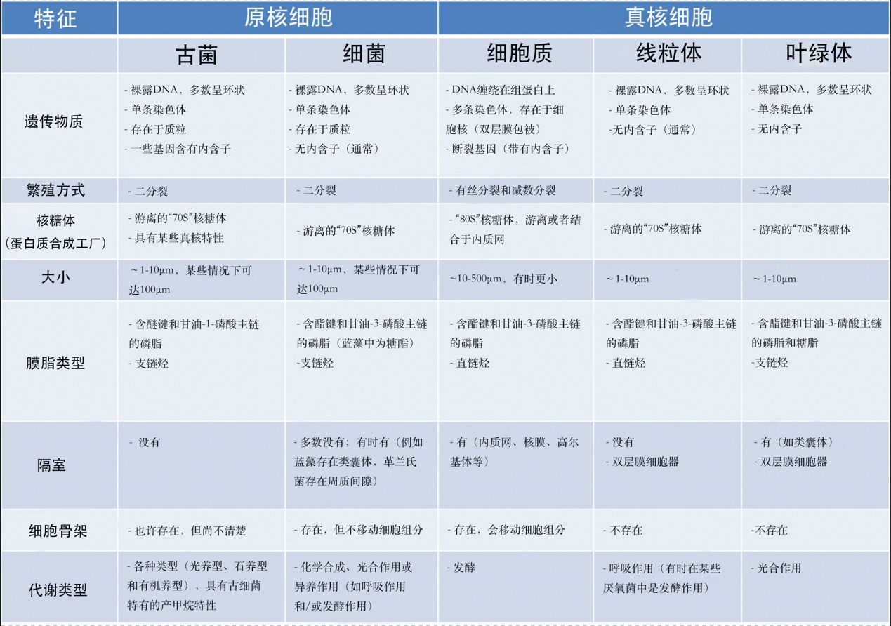
  表1比较了原核细胞和真核细胞(及其线粒体、叶绿体)的性质。说明线粒体和叶绿体具有许多与原核细胞相同的特征。除DNA结构之外,真核细胞的细胞器是由先前存在的细胞器形成的,通过分裂繁殖,就像细菌一样。同样,它们有相同的蛋白质合成机制(基质或基质中的游离70S核糖体【核糖体是由RNA和核糖体蛋白组成的庞大复合体,它使mRNA翻译成蛋白质。核糖体是所有细胞(原核生物和真核生物)共有的,但根据生物体的不同而有所变化:真核生物中为80S核糖体,原核生物和细胞器(线粒体、叶绿体)中为70S核糖体。】),而在真核细胞的细胞质中,这种机制由80S核糖体组成,有时固定在内质网膜上【内质网是真核细胞胞质中的膜网络,对细胞新陈代谢(脂质和蛋白质合成、钙储存)至关重要。与核糖体结合,是细胞内合成分泌到细胞外的蛋白质以及构成细胞器膜的蛋白质和脂质(高尔基体、溶酶体、线粒体、细胞核、核糖体、囊泡等)的地方。】。最后,细菌也有线粒体的代谢作用(即呼吸作用),在某些特殊情况下,还有叶绿体的代谢作用(即光合作用)。另一方面,真核细胞的特点是存在一个活性蛋白质网络,即细胞骨架,一个能够移动的自组织系统,负责定位和移动细胞中的细胞器。在原核生物中,这种蛋白质网络是静态的,甚至不存在的,在线粒体和叶绿体中也几乎没有这种骨架。

环境百科全书-生命-用核糖体小亚基基因构建的三个生物类群的无根系统发育树
图2. 用核糖体小亚基基因构建的三个生物类群的无根系统发育树。
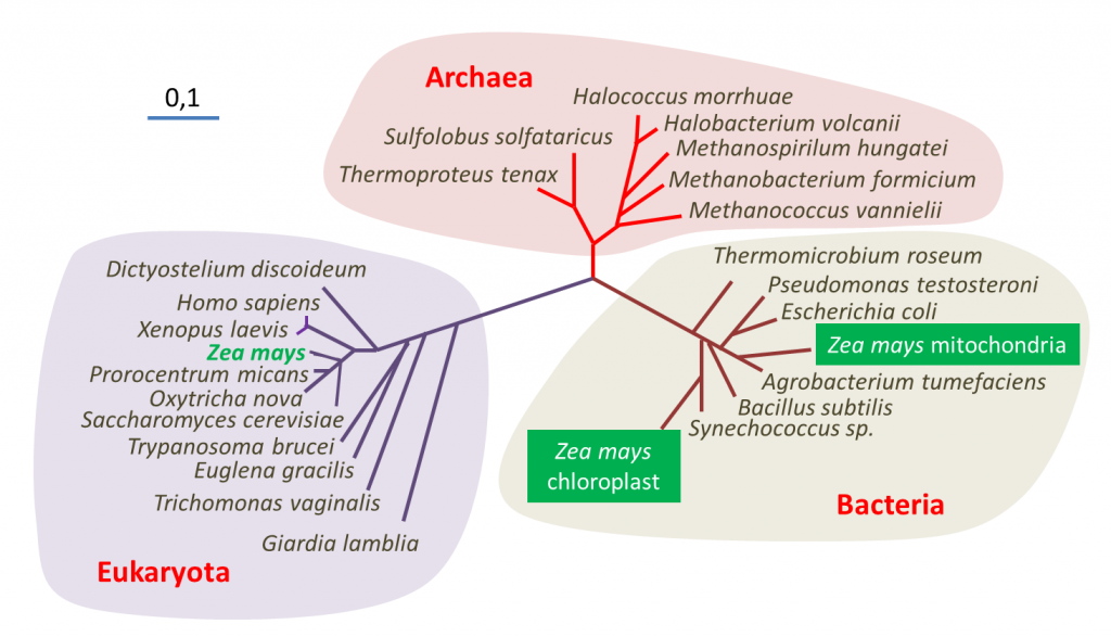
表明了玉米(Zea mays)中包含的三个基因组(细胞核、线粒体和叶绿体)的位置——聚球藻(Synechococcus)是一种蓝藻[来源:改编自参考文献[1](图2 Archaea 古菌;Bacteria 细菌;Eukaryote 真核生物;Halococcus morrhuae 嗜盐球菌属 嗜盐球菌;Halobacterium volcanii 嗜盐杆菌属 火山盐杆菌;Methanospirillum hungatei 甲烷螺菌属 亨氏甲烷螺菌;Methanobacterium formicium 甲烷杆菌属 甲酸甲烷杆菌;Methanococcus vannielii 甲烷球菌属 万氏甲烷球菌;Sulfolobus solfataricus 硫化叶菌属 硫磺矿硫化叶菌;Thermoproteus tenax 热变形菌属 附着热变形菌;Thermomicrobium roseum 热微菌属 玫瑰色热微菌;Pseudomonas testosterone 假单胞菌属 睾丸酮假单胞菌;Escherichia coli 埃希氏菌属 大肠杆菌;Zea mays mitochondria 玉米线粒体;Zea mays chloroplast 玉米叶绿体;Agrobacterium tumefaciens 农杆菌属 根癌农杆菌;Bacillus subtilis 芽孢杆菌属 枯草芽孢杆菌;Synechococcus sp. 蓝细菌聚球藻属 聚球藻;Dictyostelium discoideum 网柄菌属 盘基网柄菌;Homo sapiens 人属 智人;Xenopus laevis 爪蟾属 非洲爪蟾;Zea mays 玉米;Prorocentrum micans 原甲藻属 海洋原甲藻;Oxytricha nova 尖毛虫属 尖毛虫;Saccharomyces cerevisiae 酵母菌属 酿酒酵母;Trypanosoma brucei 锥虫属 布氏锥虫;Euglena gracilis 裸藻属 纤细裸藻;Trichomonas vaginalis 毛滴虫属 阴道毛滴虫;Giardia lamblia 贾第虫属 蓝氏贾第鞭毛虫)

  利用DNA测序技术进行基因组序列分析提供了关于生物进化史的信息,特别是关于它们的亲缘关系,也称为系统发育【研究相关物种之间联系的学科称为“系统发育学”(phylogenetics)。它允许追踪生物从共同祖先演化的主要阶段,并建立生物之间的亲缘关系。】(参阅 什么是生物多样性?以及遗传还是趋同?)。对玉米核基因组及其线粒体或叶绿体基因组进行分子系统发育分析,使确定这种植物在生命树中的系统发育位置成为可能(图2)。分析表明,三个谱系(其中两个属于细菌)在结构和功能上纠缠在一起,被认为是一个单一的有机体——显示了该物种的三重起源。

  所有这些特性表明,真核细胞是一种嵌合体,它既包含真核细胞的特征成分(细胞核),又包含具有典型原核特性的细胞器(叶绿体、线粒体)。

  1925年,爱德华·查顿(Edouard Chatton)[2] (他命名了这两种细胞类型)提出了原核细胞和真核细胞之间的区别,然而这两种细胞类型的说法直到20世纪50-60年代才被接受。自19-20世纪之交,人们观察到了真核细胞的嵌合性。1883年,法国植物学家安德烈亚斯·辛珀(Andreas Schimper)提出光合作用是不同生物体结合的结果,随后俄罗斯生物学家康斯坦丁·梅列什科夫斯基(Constantin Mereschkowsky)给出了确凿论据,即某些细胞来自两种不同类型细胞的结合(内共生)。梅列什科夫斯基(Mereschkowsky)在其1905年的文章[3]中提出了三个基本观点:(a)叶绿体是一种蓝细菌,在进化的早期就与异养宿主建立了共生关系,(b)宿主本身也是更大的异养宿主间早期共生的产物,一种较小的微球菌型内共生体形成了细胞核,以及(c)植物的自养能力完全从蓝细菌继承而来。梅列什科夫斯基没有考虑线粒体的起源。这要归功于法国微生物学家保罗·波蒂埃(Paul Portier),他在1918年的一篇文章[4]中写道:“所有生物,包括所有动植物,都是由两种不同生物结合构成的。每一个活细胞都包含一种特殊结构,细胞学家称之为“线粒体”’。 对我来说,这些细胞器只不过是共生细菌,我称之为共生体。”这些观察结果没有引起科学家们的更多关注,这一理论因此失宠,尤其是因为质体和线粒体的培养实验(在19世纪,这一实验被认为是证明质体和线粒体是细菌起源的证据[5])失败了。1970年左右,美国微生物学家林恩·马古利斯运用电子显微镜、生物化学和分子生物学等新的细胞研究方法,提出了真核细胞细胞器内共生起源的理论。

2.真核细胞是如何演化的?

环境百科全书-生命-真核生物起源假说
图3. 真核生物起源假说。
(A)一种“三界”系统,其中真核谱系和古菌谱系有相同的起源,两个谱系一样古老。(B)一种“二界”系统,来自最近的系统发育分析。
(图3 Bacteria 细菌;Archaea 古菌;Eukaryote 真核生物;Endosymbiotic organelles 内共生细胞器;Host cell for endosymbiont mitochondrial 内共生线粒体宿主细胞)

  人们提出了多种假说来解释真核细胞的出现(约15亿年前,也就是在第一批原核生物出现近10亿年后)。这个问题可以用多种方式来解释,这取决于是否综合考虑了古生物学证据、能量方面、真核细胞特征的起源或不同原核和真核系之间的相互关系[6]。图3展示了真核生物和其他谱系(古菌和细菌)的起源。

  一些模型认为,真核生物是由一个祖先谱系在进化期间经过连续突变产生的。另一些模型则认为,真核生物是由原核细胞的共生组合产生的,而这种融合导致了原核生物向真核生物过渡。这些不同的假说可以通过实验来部分验证,特别是通过对现有生物(原核或真核)基因组的分析[7,8]

环境百科全书-生命-真核细胞
图4. 古菌宿主细胞与α-变形菌融合形成含有线粒体的真核细胞[见参考文献5]。
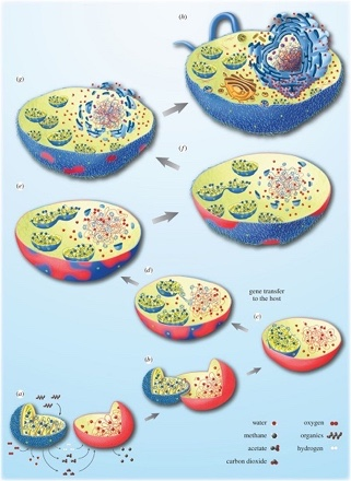
(a-h)描述从依赖氢气的古菌宿主细胞(红色)到兼性厌氧a-变形菌(蓝色),再转变为真核细胞的各种步骤。最初,这两种生物都生活在附近,古菌需要细菌产生的氢,而细菌无法承受其自身代谢产生的氢气积累。于是细菌的融合随之而来,这种转变伴随着两个生物体之间的基因转移(c),同时伴随着细胞核的建立(e-h)。[来源:经作者许可转载(见参考文献[5]) © 2015 (CC BY 4.0)](图4 water 水;methane 甲烷;acetate 醋酸酯;carbon dioxide 二氧化碳;oxygen 氧气;organics 有机物;hydrogen 氢;gene transfer to the host 基因向宿主转移)
  原始宿主细胞的性质是一个备受争议的问题。古菌是真核细胞出现的可能原因之一:在这种情况下,古菌宿主细胞和α-变形菌(属于细菌中的一大类——革兰氏阴性菌,因其细胞壁脂多糖含量高而肽聚糖含量低而得名。目前真核生物细胞中的线粒体被认为起源于这类细菌之一,即α-变形菌。)建立了稳定的共生关系(图4,[5])。如何建立这种关系存在诸多可能性[6],其中之一是:两个生活在一起的厌氧原核生物间存在营养关联,它们从这种关联中获取食物(共营养),这便表明了线粒体共生的起源。在这个假说中[5],宿主细胞是一种依赖于环境中氢气的古菌(所谓依赖氢的古菌,能够产生甲烷),共生体是一种兼性厌氧生物(α-变形菌),它既可以在有氧条件下呼吸,也可以在无氧条件下进行产氢发酵。后一种代谢只能在低浓度氢气条件下产生能量,并受益于依赖氢的古菌的存在。图4展示了这种情况下如何演变成真核细胞[5][9]。这一假说的优势在于,共生伙伴是相互需要的,而当前生物圈中存在的古菌和细菌正属于这种情况。

  在这种背景下,细菌和古菌之间密切互动,其互动形式与现今的许多共生关系相同。一般来说,这可能导致图4所示的情况,即细菌共生体将被原始宿主保留并最终驻留在宿主体内。在这种情况下,宿主并不是以共生体为食,因此融合不是由于吞噬现象造成的——尽管吞噬作用无疑增加了真核细胞进化过程中内共生体融合的频率 [5]

  真核细胞的起源也与细胞核有关,细胞核是真核细胞的标志性结构。线粒体形成后,宿主体内诞生了一种新的膜系统,即核膜,这可能是由于细菌脂质组成的膜囊聚集导致的。伴随着宿主和共生体间的基因转移,这种细胞核与细胞质的分离使得RNA剪接与DNA翻译过程分开。在这种情况下,选择压力可能导致了新形成的细胞核和细胞质之间区室化的固定[5](图4)。

  因此,目前已知的所有真核生物都来自一个古老的祖先,它在前寒武纪时期融合了一种变形菌,即线粒体。这一步是至关重要的:这次融合与真核细胞的起源密不可分。原核细胞内的能量是匮乏的,这也是驱动这种细胞进化的一个主要因素:只有具有线粒体的细胞才有足够的能量实现真核细胞的复杂性,这就是为什么没有原核生物到真核生物的中间过渡态。通常认为,只有在线粒体融合完成之后,才有部分细胞获得了真核细胞的特征(细胞核、区室化),并且在某些特殊情况下,融合蓝细菌,这就是叶绿体的起源。同时,它们获得了进行光合作用的能力,因此能够自养代谢。

  最近,一些研究揭示了线粒体共生的时间[10],他们是依据真核细胞核中存在的细菌基因的时间(即真核细胞核与游离细菌中发现的同源基因分离的日期)。他们发现,许多基因,包括一些导致真核细胞复杂性的基因,很可能是在线粒体形成之前获得的。这并不意味着线粒体进入的细胞像现在这样复杂,但它可能已经能够吞噬细胞。这种特性(真核生物特有的,依赖于细胞骨架的流动性)可能有助于线粒体的建立。因此,关于真核生物复杂性的演化仍然是推测性的,但可能在线粒体形成前就已经开始了。毫无疑问的是,线粒体形成进一步加速了这种复杂性的演化。

3.叶绿体的内共生起源

  在白细胞或许多原生动物(图5)的吞噬过程中,被摄入的细胞通常被直接消化(如猎物),但有时它们会永久滞留在细胞中(内共生体)。因此,在内共生过程中,细胞器是通过吞噬作用内化而产生的,而不是在真核生物内消化原核生物的结果(图5)。陆生植物中的叶绿体是如此,与之相近的红藻和绿藻也是如此[11,12]

环境百科全书-生命-吞噬作用与初级内共生
图5.吞噬作用与初级内共生。
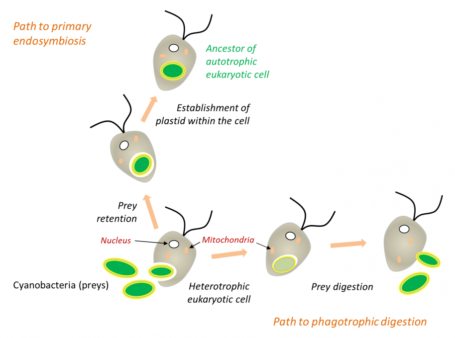
在吞噬过程中,被摄取的猎物通常被直接消化,但有时可能永久滞留在细胞中。细胞质膜包覆在原核生物周围,并将其隔离在内吞囊泡内。然后,当原核生物被融合到真核细胞中时,囊泡的膜消失了,位于蓝细菌两层膜之间的肽聚糖层也消失了[见参考文献 [9][10]
(图5 Path to primary endosymbiosis 初级内共生途径;Path to phagotrophic digestion 吞噬消化途径;Heterotrophic eukaryotic cell 异养真核细胞;Cyanobacteria (preys) 蓝细菌(猎物);Nucleus 细胞核;Mitochondria 线粒体;Prey digestion 猎物消化;Prey retention 猎物滞留;Establishment of plastid within the cell 细胞内质体的形成;Ancestor of autotrophic eukaryotic cell 自养真核细胞的祖先)
  在吞噬过程中,细胞的质膜逐渐包围猎物,将其隔离到内吞囊泡中,当这些囊泡与溶酶体(其中含有酶)囊泡融合时,就会开始消化猎物。以此类推,一般认为细胞器的外膜来自这种内吞膜。然而,事情可能更加复杂(图5)。事实上,形成叶绿体或线粒体的原核生物是革兰氏菌,这种革兰氏菌本身就存在双层膜结构。叶绿体的外膜,特别是浸没在细胞胞浆中的外表面,含有蓝细菌特有的糖脂[9][10][13]。因此,在原核生物整合到真核细胞的过程中,内吞膜可能已经消失了。目前研究发现一种特殊生物:绿叶海蛞蝓Elysia chlorotica(参阅 焦点),它是一种海洋软体动物,以藻类为食,消化藻类组织但能够长期保留其叶绿体。因此,叶绿体被融合到这种生物的细胞质中,并在整个生命周期中保持功能,这使得绿叶海蛞蝓能够像植物一样利用光合作用来获取能量生存。

  初级和次级内共生

  在进化过程中,内共生事件反复发生,导致特定生物的形成。在初级内共生过程中,真核细胞融合活的原核生物。因此,绿系植物(例如属于陆生植物的红藻和绿藻)的叶绿体源自含有蓝细菌的初级内共生体。在一些真核生物中,线粒体因适应厌氧环境而演化,但从未消失:它们产生特定的线粒体,如氢化酶体(hydrogenosomes),进行产氢发酵(例如某些纤毛虫)[14],但也产生小的细胞器,只参与宿主细胞的生物合成,如纺锤剩体(mitosomes)[15]

环境百科全书-生命-共生模型
图6. 蓝隐藻(Guillardia theta)的次级叶绿体内共生模型[见参考文献[18]]。
在这种情况下,红藻细胞核(初级宿主)以退化的细胞核(即核形体)的形式存在于次级宿主中,但基因组非常小(仅551 kb)。叶绿体和线粒体的基因组也大大减少。(图6 Primary endosymbiosis 初级内共生;Primary eukaryotic cell 初级真核细胞;Secondary endosymbiosis 次级内共生;Nucleus 细胞核;Cyanobacteria 蓝细菌;Mitochondrion 线粒体;Cryptomonad cell 隐滴虫细胞;Chloroplast 叶绿体;Nucleomorph 核形体)
  二次内共生是初级内共生过程的重复,当一个已经包含一个内共生体的真核生物在另一个真核生物中实现了一个次级内共生(图6)。这是多膜(超过两层膜)质体的起源:如眼虫属(Euglena)中绿藻的内化;褐藻中红藻的独立内化等。有时还存在三次内共生,比较少见,但也被描述过。这些不同的共生体构成了进化谱系的原始内共生体 [16,17]

4.原核生物与真核细胞的融合

  所有这些品系都有一个共同的特点:内共生体存在强烈遗传退化。与大肠杆菌(Escherichia coli)等游离变形菌相比,线粒体丢失了99%的基因。在极端情况下,氢化酶体(hydrogenosomes)和纺锤剩体(mitosomes)根本没有基因组!与单细胞游离蓝细菌相比,绿系质体显示出95%的遗传退化:蓝细菌的基因数量从几千个降低到叶绿体中的100到200个左右……寄生植物大花草属(Rafflesia)中的质体甚至没有基因组。

  这种退化显然是因为缺乏自由生活甚至是某些代谢功能所必需的基因。例如,与所有革兰氏菌【革兰氏菌是通过一种名为革兰氏染色的染色技术检测到的细菌:它们在显微镜下呈现粉红色。染色技术基于细菌的细胞膜和细胞壁的特征。然而,这并不是一个系统发育分类因素:事实上,“革兰氏阳性”组和“革兰氏阴性”组都是非单系的。革兰氏阳性和革兰氏阴性细菌细胞壁由一层肽聚糖构成,肽聚糖的主要结构可以分为两部分:多糖部分和肽部分。这层肽聚糖保持细胞的形状并保护细胞免受渗透压的影响】一样,蓝细菌的两层膜间有一层肽聚糖,这对于在低渗透压【渗透压指每升溶液中具有“渗透活性”的粒子的摩尔数。是一个与溶液中粒子施加的渗透压相关的概念,与渗透相关。蔗糖是一种小型的渗透活性分子,而淀粉是一个巨大的渗透不活性葡萄糖聚合物。在一个区室中蔗糖的积累导致该区室中的渗透压增加,而淀粉则不是这种情况。】的自然环境中维持细菌的结构至关重要。一旦原核生物融合到宿主细胞,原核生物将处于细胞质中,其渗透压非常接近原核生物自身内部介质的渗透压。显然,此时将不再需要肽聚糖层,于是叶绿体中有关肽聚糖层的基因逐渐丢失(灰胞藻除外)。

  尽管细胞器基因组正在退化,但已知的细胞器蛋白质库(蛋白质组)仍然与游离细菌蛋白质组相似:因此,这些额外的蛋白质可能弥补了基因组的缺失。它们的编码实际上由宿主核基因组负责:细胞核基因在细胞质中翻译成蛋白质,通过转运肽(一种肽序列,位于细胞质中新合成蛋白质的NH2末端,定向到特定的细胞器(线粒体等)以执行其功能,也称之为定向肽)到达细胞器。这种基因产物的重新定位,对于原核生物融合到宿主细胞是必要的。负责转移的寻址机制是质体和线粒体的融合创新,是细胞内共生体的又一个新功能。这种机制允许细胞质中合成的蛋白质,通过跨膜转运进入线粒体和叶绿体中发挥作用。这种寻址涉及大量进化起源复杂的蛋白质:原核或者真核来源;由细胞器或者细胞核编码。它们共同起作用,允许识别正在寻址的蛋白质,使其展开(蛋白质必须保持在非折叠的状态才能穿过膜),然后导入,随后切割寻址肽,最终将蛋白精确定位到其所在功能区[19]

  这种在细胞器中发挥功能的细胞核编码基因,究竟是如何起源的?实际上存在两种可能(图7)[16]。有时,原始核基因取代了细胞器基因:对应的基因产物能够精确定位到细胞器中发挥功能。然而,如果细胞器中已经存在了一个编码相同功能的基因,那么这种定位转移可能导致蛋白质的冗余。由于这种冗余,细胞器基因可能逐渐丢失而不损害其功能(图7a)[16]

环境百科全书-生命-进化机制
图7. 导致细胞器基因被位于细胞核的基因取代的进化机制。
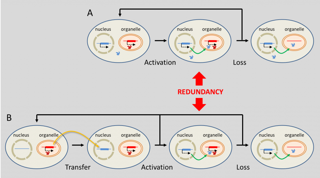
(A)“真正的”细胞核起源的基因取代了细胞器基因;(B)细胞器向细胞核方向的基因转移。[来源:根据瑟洛斯(Selosse)等人(2001年)参考文献[16]]。(图7 nucleus 细胞核;organelle 细胞器;Activation 活化;Loss 丢失;Redundancy 冗余;Transfer 转移)
  另一种情况涉及从细胞器向细胞核的基因转移,主要分为两个阶段(图7b)。首先,编码细胞器蛋白的DNA片段被重新定位,然后融合到核基因组中。通过突变适应核遗传密码,并且获得转录的调控序列,这时这段融合序列才会开始编码。此外,核基因组还必须获得与转运肽相对应的DNA序列,才能确保成熟蛋白能正确定位到细胞器中。由此一来,出现了基因冗余:任意一个副本都可能丢失。功能的丧失和/或细胞器副本的丢失,随即掩盖了这种基因转移的过程(图7)[16]

  DNA片段从细胞器转移到细胞核并不罕见:某些植物的基因组中插入了大片段的细胞器DNA。这些基因可以被激活:拟南芥中近10%的核基因是从质体转移而来的,其次是复制子[20]。目前尚不清楚内共生体的DNA是如何融合到宿主基因组中的,但人们认为,这可能发生在受损或衰老的细胞器降解过程中,DNA片段意外地释放到宿主细胞质,然后随机融合到宿主核DNA中。

  细胞器的细胞质基因组处于各种选择压力下,其中一些选择力促进它们的退化(例如某些基因的共表达需要),另一些选择力有利于特定基因在细胞器基因组中保留。细胞器小基因组选择可能就是这种情况,它加速了细胞器的繁殖,允许更好地传递遗传信息给子代细胞:例如,它选择将基因转移到细胞核。因此,它积累了来自不同谱系的遗传潜力,这些谱系与它在细胞中共存[16]。因此,虽然内共生减少了内共生体的基因组,但它滋养了宿主细胞核的基因组,促进了核基因组的遗传多样化,并推动了更紧密的内共生关系形成。因此,通过多次内共生嵌套以及宿主细胞核基因嵌合,内共生体逐渐构成了目前的进化谱系。

  最后,内共生体的跨代垂直转移对于内共生体的持续存在至关重要。质体必须在宿主细胞分裂前分裂,并且必须均匀分布在两个子细胞中。如果分裂得太快,它们可能比宿主细胞占优势。相反,分裂得太慢则可能导致内共生体消失。在这种情况下,细胞分裂和共生体分裂的协调进行是内共生成功的关键因素。虽然参与叶绿体分裂的大多数蛋白质来自蓝细菌的细胞分裂机制,但有些蛋白质显然是真核来源的(这种蛋白都在细胞核中编码):这是宿主控制叶绿体分裂的一种方式。

5.共生驱动进化吗?

环境百科全书-生命-质体进化史上的内共生
图8. 质体进化史上的内共生(初级、次级和三级)。
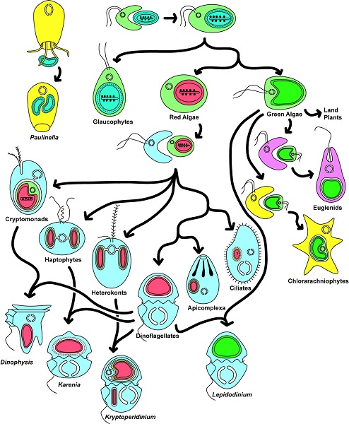
由此产生了各种各样的生物,如红藻和绿藻、陆生植物、顶复门生物(如疟原虫、弓形虫,可导致疟疾)或甲藻(一种海洋浮游生物,在初级海洋生产中尤为重要)。[来源:转载自基林(Keeling)等人[12]。 ©2016 美国植物学杂志版权所有](图8 Paulinella 阿米巴原虫;Glaucophytes 灰胞藻;Red Algae 红藻;Green Algae 绿藻;Land Plants 陆地植物;Euglena 裸藻;Chlorarachniophytes 海洋阿米巴鞭毛原虫;Cryptomonads 隐藻;Haptophytes 定鞭金藻;Heterokonts 不等鞭毛藻;Dinoflagellates 甲藻;Apicomplexa 顶复门生物;Ciliates 纤毛虫;Dinophysis 鳍藻;Karenia 凯伦藻;Kryptoperidinium 隐多甲藻;Lepidodinium 鳞甲藻)

  综上所述,这种多样化的共生关系导致了真核细胞的形成[1,9],是真核生物进化过程中多样性发展的基础。内共生是新的进化谱系形成的基础。图8展示了内共生生物的极端多样性。然而,事情远比想象的复杂:这种共生进化过程可能会持续重复!今天,一些独特的单细胞藻类,如隐藻【一种单细胞生物,多数能进行光合作用。它们的叶绿体由四个膜包围,表明它与能够进行光合作用的真核生物进行了内共生。隐藻存在于许多环境中,特别是水生环境(海洋环境、淡水、湿地孔隙水)。一些物种已经成为后生动物的肠道寄生虫。有些是甲藻的内共生体。】和异形藻(图8),由二次内共生质体进一步融合到甲藻细胞质内形成(这种情况下,甲藻失去了原有的质体)。它们的质体含有四层膜,属于二次内共生质体:这里发生了三次连续的内共生!

  共生不仅仅是一种生物学现象,它还是推动生命世界进化的强大动力之一。它能迅速创造出嵌合生物,可以产生新的细胞系。它拉近了共生伙伴间的距离,促进了大规模的基因转移,创造了嵌合基因组:这种核基因组包含真核基因,同时也包含细菌基因(来源于线粒体,甚至与之相邻生物的质体)。这些多样的共生事件可以解释生命进化史上的重大飞跃(不连续的进化事件),它们创造了强大的生命,塑造了当前的生物多样性。

  因此,从达尔文进化观点来看,一个物种可能会产生两个物种(即所有物种来自于一个共同祖先),但内共生机制提醒我们,有时两个物种(自由生活的、独立可区分的)也可能合并成一个新的物种。人类可以被看作是一个高度融合的共生群落,由真核细胞质和线粒体(起源于原核生物)组成,同时也包括古菌和细菌(例如肠道微生物)。


参考资料和说明

[1] Lang T. et al (2000) Autophagy and the cvt pathway both depend on AUT9. J Bacteriol 182, 2125-2133.

[2] Chatton E. (1938) Titres et travaux scientifiques (1906-1937). Sette, Sottano, Italy. L’histoire des conditions dans lesquelles Chatton a établi le concept de procaryote et eucaryote est décrite par Sapp J. (2005) The Prokaryote-Eukaryote Dichotomy: Meanings and Mythology, Microbiol Mol Biol Rev. 69, 292–305.

[3] Mereschkowsky C. 1905 Uber Natur und Ursprung der Chromatophoren im Pflanzenreiche. Centralbl. 25, 593-604; translated by Martin W, Kowallik K. (1999) Annotated English translation of Mereschkowsky’s 1905 paper ‘Uber Natur und Ursprung der Chromatophoren im Pflanzenreiche‘. Eur. J. Phycol. 34, 287–295.

[4] Portier P. (1918) Les Symbiotes. Masson (ed.), Paris. (in french)

[5] Martin W.F., Garg S. & Zimorski V. (2015) Endosymbiotic theories for eukaryote origin. Phil. Trans. R. Soc. B370, 20140330.

[6] Selosse M.A. (2012). Gloire et disgrâce de la théorie endosymbiotique. La Recherche 468: 92-94. (in french)

[7] Archibald J.M. (2014) One plus one equals one: symbiosis and the evolution of complex life. Oxford, UK: Oxford University Press.

[8] McFadden G.I. (2014) Origin and Evolution of Plastids and Photosynthesis in Eukaryotes, Cold Spring Harb.Perspect. Biol. 6, a016105

[9] Martin W. & Müller M. (1998) The hydrogen hypothesis for the first eukaryote. Nature 392, 37-41.

[10] Ettema T.J.G. (2016) Mitochondria in the second act. Nature 531, 39-40doi:10.1038/nature16876

[11] Archibald J.M. & Keeling P.J. (2002) Recycled plastids: a “green movement” in eukaryotic evolution. Trends Genetics 18, 577-584.

[12] Keeling P.J. (2004) Diversity and evolutionary history of plastids and their hosts. Am. J. Bot. 91, 1481-1493.

[13] Douce R., Block M.A., Dorne A.J., Joyard J. (1984) The plastid envelope membranes: their structure, composition, and role in chloroplast biogenesis. Subcell. Biochem. 10, 1-84, Springer US (Ed.)

[14] Selosse M.A. & Loiseaux-de Goër S. (1997) La Saga de l’endosymbiose, La Recherche 296, 36 (in french)

[15] Embley T.M. & Martin W. (2006) Eukaryotic evolution, changes and challenges. Nature 440, 623-630

[16] Lefèvre T., Renaud F., Selosse M.-A. & Thomas F. (2010). Évolution des interactions entre espèces, in F. Thomas, T. Lefèvre & M. Raymond (ed.), Biologie évolutive, p. 530-613. De Boeck, Paris. (in french)

[17] Keeling P.J. (2010) The endosymbiotic origin, diversification and fate of plastids. Phil. Trans. R. Soc. B 365, 729-748

[18] Douglas S. et al (2001) The highly reduced genome of an enslaved algal nucleus. Nature 410, 1091-1096.

[19] Selosse M.A., Albert B. & Godelle B. (2001) Small is successful: selection for reducing organelle’s genome size favours gene transfer to the nucleus. Trends Ecol Evol 16, 135-141.

[20] Jarvis P. (2004) Organellar Proteomics: Chloroplasts in the Spotlight. Current Biology 14, R317-9. http://www.cell.com/current-biology/references/S0960-9822%2804%2900231-3


The Encyclopedia of the Environment by the Association des Encyclopédies de l'Environnement et de l'Énergie (www.a3e.fr), contractually linked to the University of Grenoble Alpes and Grenoble INP, and sponsored by the French Academy of Sciences.

To cite this article: SELOSSE Marc-André, JOYARD Jacques (March 12, 2024), 共生与进化:真核细胞的起源, Encyclopedia of the Environment, Accessed July 27, 2024 [online ISSN 2555-0950] url : https://www.encyclopedie-environnement.org/zh/vivant-zh/symbiosis-and-evolution-origin-eukaryotic-cell/.

The articles in the Encyclopedia of the Environment are made available under the terms of the Creative Commons BY-NC-SA license, which authorizes reproduction subject to: citing the source, not making commercial use of them, sharing identical initial conditions, reproducing at each reuse or distribution the mention of this Creative Commons BY-NC-SA license.